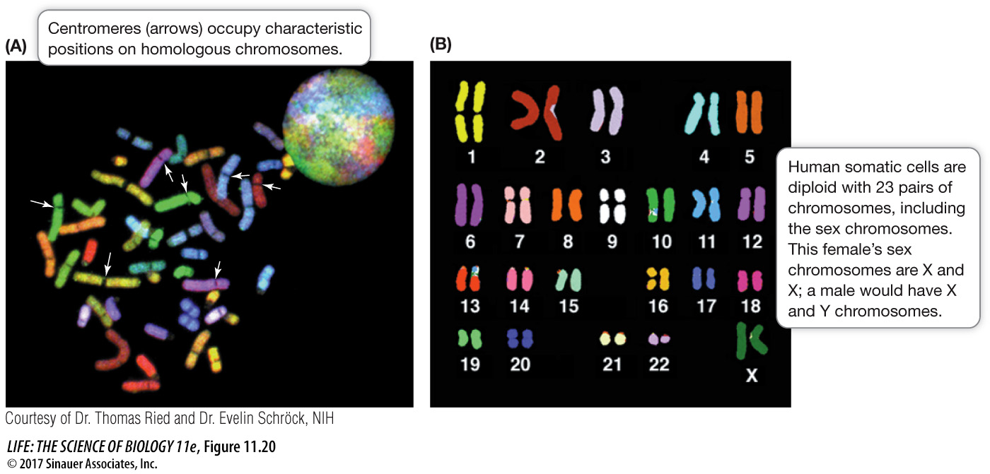
The number, shapes, and sizes of the metaphase chromosomes constitute the karyotype
When cells are in metaphase of mitosis, it is often possible to count and characterize their individual chromosomes. If a photomicrograph of the entire set of chromosomes is made, the images of the individual chromosomes can be manipulated to pair them and place them in an orderly arrangement. Such a rearranged photomicrograph reveals the number, shapes, and sizes of the chromosomes in a cell, which together constitute its karyotype (Figure 11.20). In humans, karyotypes can aid in the diagnosis of chromosomal abnormalities such as trisomies or translocations, and this has led to an entire branch of medicine called cytogenetics. However, as you will see in Chapter 15, chromosome analysis with the microscope is replaced in some cases by direct analysis of DNA.