Global chromosome changes involve DNA methylation
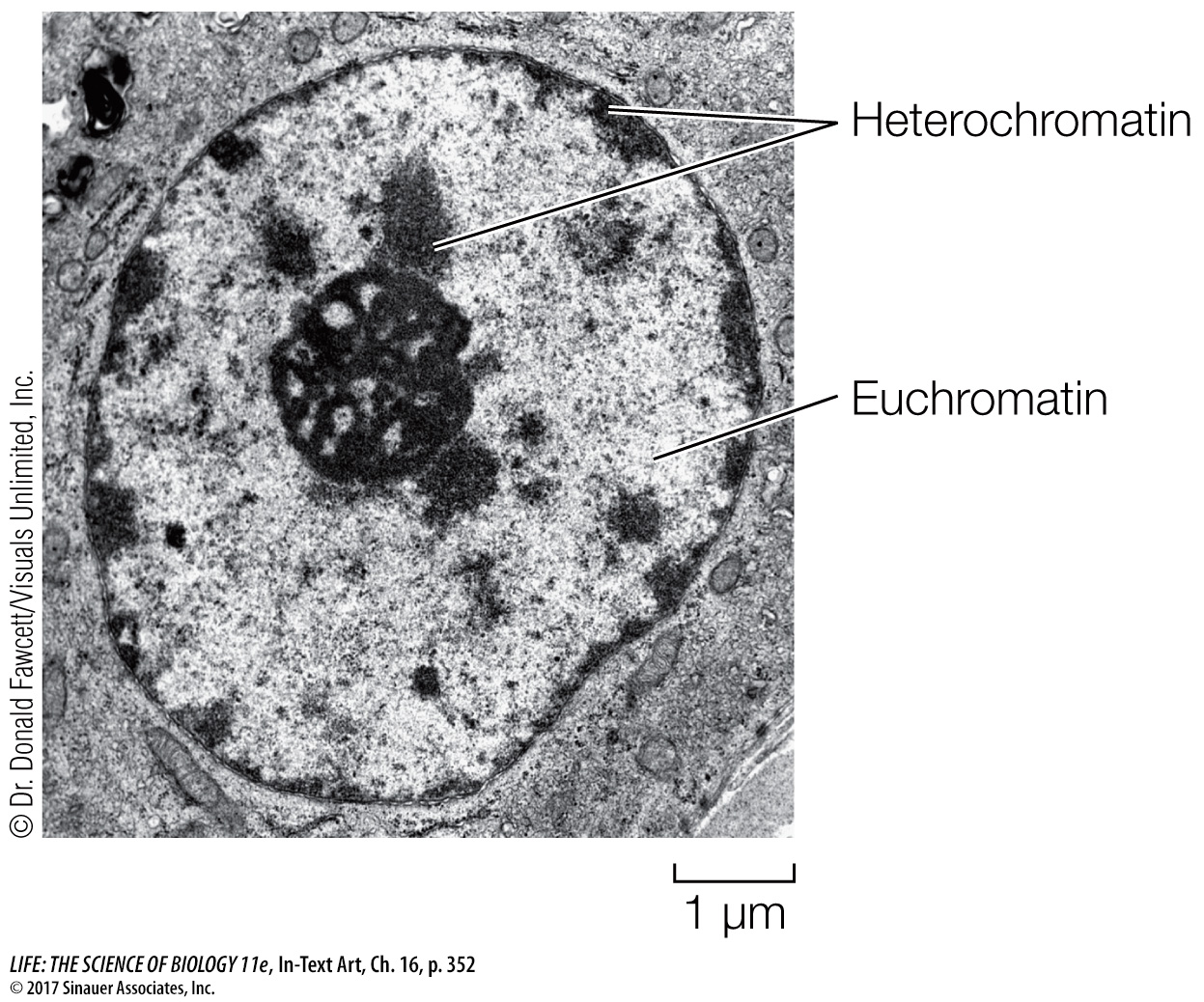
Like single genes, large regions of chromosomes or even entire chromosomes can have distinct patterns of DNA methylation. Under a microscope, two kinds of chromatin can be distinguished in the stained interphase nucleus: euchromatin and heterochromatin. The euchromatin appears diffuse and stains lightly; it contains the DNA that is transcribed into mRNA. Heterochromatin is condensed and stains darkly; any genes it contains are generally not transcribed.
Perhaps the most dramatic example of heterochromatin is the inactive X chromosome of mammals. A normal female mammal has two X chromosomes; a normal male has an X and a Y (see Key Concept 12.4). The X and Y chromosomes probably arose from a pair of autosomes (non–
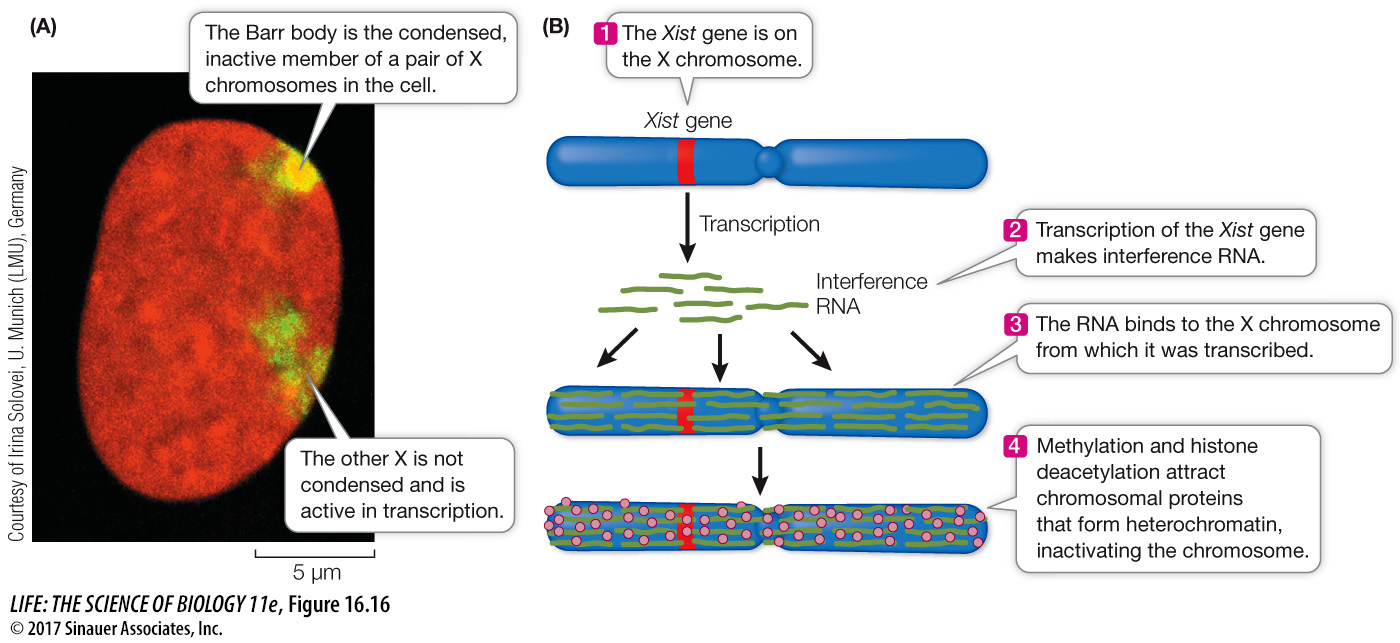
During early embryonic development, one of the X chromosomes in each cell of a female is largely inactivated with regard to transcription. The same X chromosome remains inactive in all of that cell’s descendants. In a given embryonic cell, the “choice” of which X in the pair to inactivate is random. Recall that one X in a female comes from her father and one from her mother. Thus in one embryonic cell the paternal X might be the one remaining transcriptionally active, but in a neighboring cell the maternal X might be active.
The inactivated X chromosome is identifiable within the nucleus because it is very compact, even during interphase. Typically, a nuclear structure called a Barr body (after its discoverer, Murray Barr) can be seen in human female cells under the light microscope (Figure 16.16A). This clump of heterochromatin, which is not present in normal males, is the inactivated X chromosome, and it consists of heavily methylated DNA. A female with the normal two X chromosomes will have one Barr body, whereas a rare female with three Xs will have two, and an XXXX female will have three. Males that are XXY will have one. These observations suggest that the interphase cells of each person, male or female, have a single active X chromosome, and thus a constant dosage of expressed X chromosome genes.
353
Condensation of the inactive X chromosome makes its DNA sequences physically unavailable to the transcriptional machinery. Most of the genes of the inactive X are heavily methylated. However, one gene, Xist (for X inactivation-