Alveolates have sacs under their cell membranes
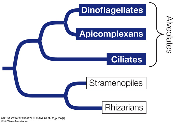
Alveolates are so named because they possess sacs, called alveoli, just beneath their cell membranes, which may play a role in supporting the cell surface. All alveolates are unicellular, and most are photosynthetic, but they are diverse in body form. The alveolate groups we will consider in detail here are the dinoflagellates, apicomplexans, and ciliates.
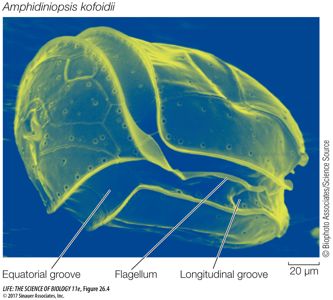
DINOFLAGELLATES Most dinoflagellates are marine and photosynthetic; they are important primary producers of organic matter in the oceans. Although fewer species of dinoflagellates live in fresh water, individuals can be abundant in freshwater environments. The dinoflagellates are of great ecological, evolutionary, and morphological interest. A distinctive mixture of photosynthetic and accessory pigments gives their chloroplasts a golden brown color. Some dinoflagellate species cause red tides, as discussed at the start of this chapter. Other species are photosynthetic endosymbionts that live within the cells of other organisms, including invertebrate animals (such as corals; see Investigating Life: Can Corals Reacquire Dinoflagellate Endosymbionts Lost to Bleaching?) and other marine protists (see Figure 26.12A). Still others are nonphotosynthetic and live as parasites within other marine organisms.
557
Dinoflagellates have a distinctive appearance. They generally have two flagella, one in an equatorial groove around the cell, the other starting near the same point as the first and passing down a longitudinal groove before extending into the surrounding medium (Figure 26.4). Some dinoflagellates can take on different forms, including amoeboid ones, depending on environmental conditions. It has been claimed that the dinoflagellate Pfiesteria piscicida can occur in at least two dozen distinct forms, although this claim is highly controversial. In any case, this remarkable dinoflagellate, when present in large enough numbers, is harmful to fish and can both stun and feed on them.
Media Clip 26.1 A Dinoflagellate Shows Off Its Flagellum
www.life11e.com/
APICOMPLEXANS The exclusively parasitic apicomplexans derive their name from the apical complex, a mass of organelles contained in the apical end (the tip) of the cell. These organelles help the apicomplexan invade its host’s tissues. For example, the apical complex enables Plasmodium, the causative agent of malaria, to enter its target cells in the human body after transmission by a mosquito.
Like many obligate parasites, apicomplexans have elaborate life cycles featuring asexual and sexual reproduction through a series of very dissimilar life stages (see Figure 26.20). In many species, these life stages are associated with two different types of host organisms, as is the case with Plasmodium. Another apicomplexan, Toxoplasma, alternates between cats and rats to complete its life cycle. A rat infected with Toxoplasma loses its fear of cats, which makes it more likely to be eaten by, and thus transfer the parasite to, a cat.
558
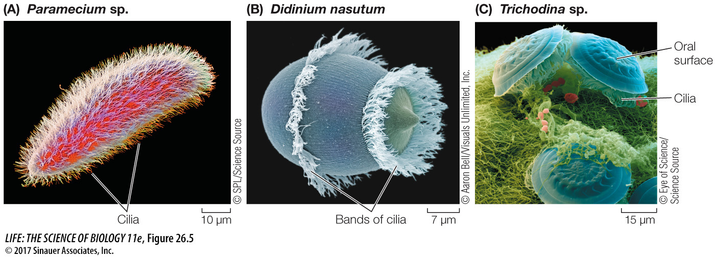
CILIATES The ciliates are named for their numerous hairlike cilia, which are shorter than, but otherwise identical to, eukaryotic flagella. The ciliates are much more complex in body form than are most other unicellular eukaryotes (Figure 26.5). Their definitive characteristic is the possession of two types of nuclei (whose roles we will describe in Key Concept 26.3 when we discuss protist reproduction). Almost all ciliates are heterotrophic, although a few contain photosynthetic endosymbionts.
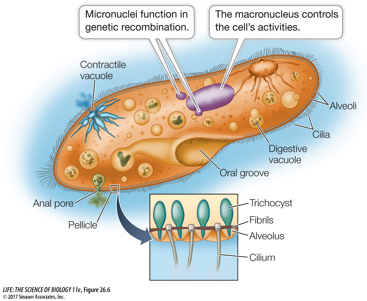
Paramecium, a frequently studied ciliate genus, exemplifies the complex structure and behavior of ciliates (Figure 26.6). The slipper-
Activity 26.1 Anatomy of Paramecium
www.life11e.com/
The cilia provide Paramecium with a form of locomotion that is generally more precise than locomotion by flagella. A Paramecium can coordinate the beating of its cilia to propel itself either forward or backward in a spiraling manner. It can also back off swiftly when it encounters a barrier or a negative stimulus. The coordination of ciliary beating is probably the result of a differential distribution of ion channels in the cell membrane near the two ends of the cell.
Organisms living in fresh water are hypertonic to their environment. Many freshwater protists, including Paramecium, address this problem by means of specialized contractile vacuoles that excrete the excess water the organisms constantly take in by osmosis. The excess water collects in the contractile vacuoles, which then contract and expel the water from the cell.
Paramecium and many other protists engulf solid food by endocytosis, forming a digestive vacuole within which the food is digested (Figure 26.7). Smaller vesicles containing digested food pinch away from the digestive vacuole and enter the cytoplasm. These tiny vesicles provide a large surface area across which the products of digestion can be absorbed by the rest of the cell.
experiment
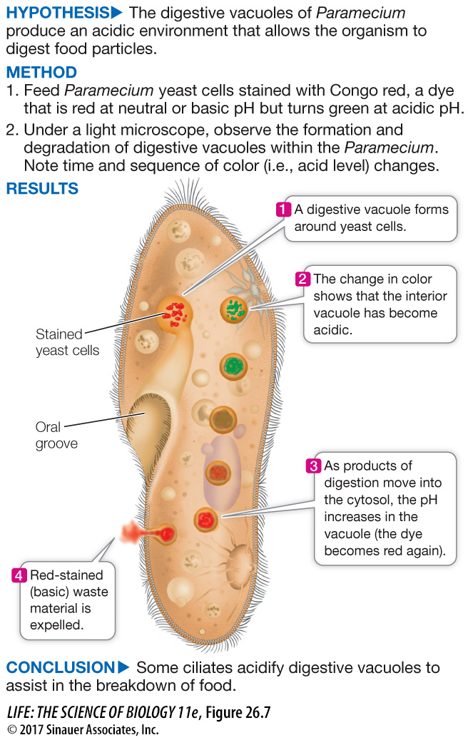
Figure 26.7 The Role of Vacuoles in Ciliate Digestion
Original Paper: Mast, S. O. 1947. The food-
An acidic environment is known to aid digestion in many multicellular organisms. Do ciliates also use acid to obtain nutrients?
Animation 26.2 Digestive Vacuoles
www.life11e.com/