The products of the stem’s primary meristems become stem tissues
Now let’s turn to the shoot, which comprises the stem and leaves. As seen in Figure 33.1, shoots and roots are composed of repeating modules called phytomers. A phytomer in the shoot system consists of a node with its attached leaf or leaves, the internode below the node, and axillary buds, each of which forms in the angle between a leaf and the stem. Shoots grow by adding new phytomers. Initially these form only on the primary stem of the plant, but if an axillary bud develops into a branch, it does so by producing new phytomers. The new phytomers originate from cells in the shoot apical meristems, which are present in the terminal buds of each branch and the main stem (see Figure 33.9).
The shoot apical meristem, like the root apical meristem, forms three primary meristems: protoderm, ground meristem, and procambium. These primary meristems, in turn, give rise to the three shoot tissue systems.
As the shoot grows and extends, bulges called leaf primordia develop on the sides of the shoot apical meristem at regular intervals (see Figure 33.9). These primordia are made up of primary meristematic tissues, which go on to develop into the mature tissues of the leaf. In addition, bud primordia form at the bases of the leaf primordia. These have the potential to become new apical meristems and initiate new shoots. The sites where leaf primordia form become nodes on the developing stem. The regions between the nodes (the internodes) lengthen initially via cell division in the primary meristematic tissues, and later by cell elongation in the mature stem tissues. The growing stem has no protective structure analogous to the root cap, but the leaf primordia can act as a protective covering for the shoot apical meristem.
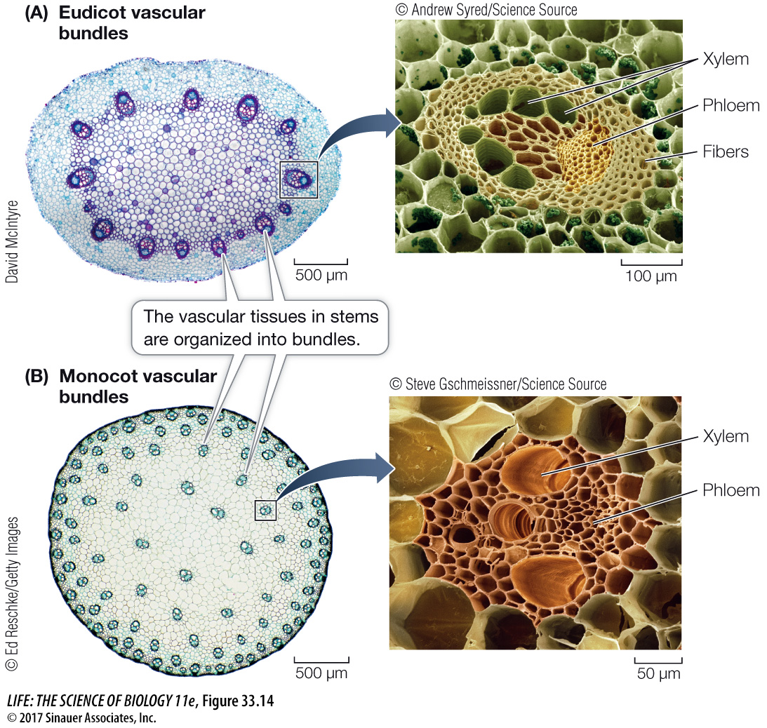
The vascular systems of stems differ from those of roots. In a root, the vascular tissue lies deep in the interior, with the xylem at or near the center (see Figure 33.11). The vascular tissue of a young stem, however, is divided into discrete vascular bundles (Figure 33.14). Each vascular bundle contains both xylem and phloem. In eudicots the vascular bundles generally form a ring, whereas in monocots they are scattered throughout the stem.
Activity 33.3 Eudicot Stem
www.life11e.com/
Activity 33.4 Monocot Stem
www.life11e.com/
In addition to the vascular tissues, the stem contains other important storage and supportive tissues. In eudicots the pith lies inside the ring of vascular bundles and also extends between them, forming regions called pith rays. To the outside lies the cortex, which may contain supportive collenchyma cells with thickened walls. The pith and cortex constitute the ground tissue system of the stem. The outermost cell layer of the young stem is the epidermis.