The pituitary is an interface between the nervous and endocrine systems
The pituitary gland sits in a depression at the bottom of the skull, just over the back of the roof of the mouth (Figure 40.6). It is attached by a stalk to the hypothalamus, which is involved in many physiological regulatory systems such as that of thermoregulation (see Key Concept 39.5). Through its close connection with the hypothalamus, the pituitary serves as the interface between the nervous system and the endocrine system and is involved in the hormonal control of many physiological processes.
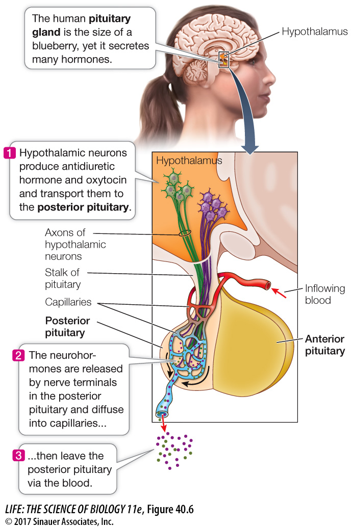
The pituitary has two parts with different developmental origins. The anterior pituitary originates as an outpocketing of the roof of the embryonic mouth cavity. The posterior pituitary originates as an outpocketing of the floor of the developing brain. Thus the anterior pituitary originates from gut epithelial tissue and the posterior pituitary from neural tissue. Both parts interact with the nervous system but in different ways. The anterior pituitary is controlled by hypothalamic neurohormones that reach the anterior pituitary via the blood. The posterior pituitary contains long extensions of hypothalamic neurons that release their neurohormones in the posterior pituitary.
Animation 40.1 The Hypothalamic–
www.life11e.com/
THE POSTERIOR PITUITARY The long hypothalamic neuron extensions into the posterior pituitary are called axons. The terminals of these axons in the posterior pituitary contain vesicles of neurohormones—
*connect the concepts As described in Key Concept 45.2, action potentials are sudden and transient electric signals generated by voltage-
The main action of antidiuretic hormone (ADH) in mammals and birds is to increase the amount of water conserved by the kidneys. When ADH secretion is high, the kidneys produce only a small volume of highly concentrated urine. When ADH secretion is low, the kidneys produce a large volume of dilute urine. The posterior pituitary increases its release of ADH when blood pressure falls or the blood becomes too salty. ADH is also known as vasopressin because at high concentrations it causes the constriction of peripheral blood vessels as a means of elevating blood pressure.
851
The hormone oxytocin is released from the posterior pituitary when a woman is about to give birth. Oxytocin stimulates contractions of the uterine muscles, resulting in the delivery of the baby. Oxytocin also brings about the flow of milk from the mother’s breasts. The baby’s suckling stimulates neurons in the mother’s brain that cause the secretion of oxytocin. Even the crying of her baby can cause a nursing mother to secrete oxytocin, resulting in the release of breast milk—
THE ANTERIOR PITUITARY The anterior pituitary produces and releases into the circulation four peptide and protein hormones that act as tropic hormones, meaning they control the activities of other endocrine glands. These four tropic hormones are thyrotropin (thyroid-
Growth hormone (GH) acts on a wide variety of tissues to promote growth and development. One of its important effects is to stimulate cells to take up amino acids. Growth hormone also promotes growth by stimulating the liver to produce chemical signals that stimulate the growth of bone and cartilage. Overproduction of GH in children causes gigantism; affected individuals may grow to nearly 8 feet tall. Underproduction of GH results in pituitary dwarfism, in which individuals fail to reach normal adult height.
Endorphins and enkephalins are the body’s natural painkillers. In the brain, these molecules act as neurotransmitters in pathways that control pain. Their production in the anterior pituitary is normally quite small and probably has little significant effect.
Melanocyte-