Hormones influence the nervous system
Hormones have more influences on the nervous system than simply being negative feedback signals controlling their own secretion. For example, the posterior pituitary hormone oxytocin, which plays a role in the birth process and in stimulating the flow of milk from the breasts, also has a strong behavioral effect in promoting bonding (see the story that opens Chapter 7). If oxytocin release is experimentally blocked, mammalian mothers, from rats to sheep, will reject their newborn offspring, but if a virgin rat is given a dose of oxytocin, she will adopt strange pups as if they were her own. Oxytocin promotes pair bonding in a variety of animals. In humans, oxytocin secretion rises with intimate sexual contact, and it has been nicknamed the “cuddle hormone.” Experiments using games involving financial exchanges have shown that puffs of oxytocin applied to the nostrils raise the level of trust between players. Thus circulating hormones can have a strong influence on behavior. Another example is the effects of sex hormones, which we will discuss in Key Concept 53.3.
852
Recent investigations involving the hormone irisin that was presented at the beginning of this chapter reveal that this same molecule may mediate effects of exercise on the brain. Many experiments have shown that exercise improves cognitive functions. Beneficial effects of exercise have been shown especially for a part of the brain called the hippocampus that is involved in learning and memory. These effects include the production of new brain cells, increased blood flow, and changes in the structures supporting communication between brain cells. It had been established that one molecular factor involved in the beneficial effects of exercise on the hippocampus is BDNF, which stands for brain-
853
investigating life
How Could Irisin Mediate the Benefits of Exercise on Cognition?
experiment
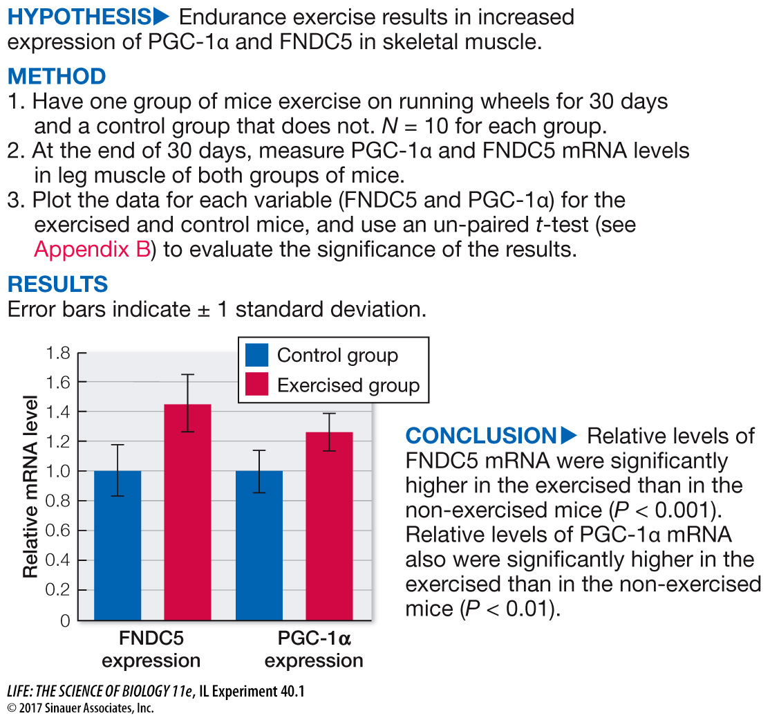
Original Paper: Wrann, C. D. et al. 2013. Exercise induces hippocampal BDNF through a PGC-
Irisin secreted by exercising muscle is cleaved from a protein called FNDC5, and FNDC5 expression is controlled by the transcription factor PGC-
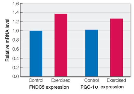
work with the data
Similar measurements of expression of FNDC5 and PGC-
| FNDC5 | PGC- |
||
|---|---|---|---|
| Control | Exercised | Control | Exercised |
| 1.02 | 1.33 | 1.22 | 1.23 |
| 1.10 | 1.24 | 0.84 | 1.09 |
| 0.92 | 1.68 | 1.06 | 1.35 |
| 0.93 | 1.30 | 1.26 | 1.18 |
| 1.21 | 1.34 | 1.04 | 1.40 |
| 0.93 | 1.33 | 1.12 | 1.18 |
| 0.96 | 1.50 | 1.04 | 1.46 |
| 1.01 | 1.29 | 1.09 | 1.19 |
| 1.00 | 1.44 | 1.02 | 1.54 |
| 0.97 | 1.41 | 0.54 | 1.16 |
QUESTIONS
Question 1
Plot the data as was done for the leg skeletal muscle results. Then you can apply the same analysis methods to these data that were applied to the leg muscle data in the preceding experiment. Did exercise have a significant effect on FNDC5 and PGC-
Question 2
The neurotrophic factor BDNF is a secreted protein that plays important roles in neural changes associated with learning and memory. How would you determine if the exercise protocol induced a significant increase in BDNF expression in the hippocampus, assuming you have an assay for BDNF?
To determine if exercise increases BDNF levels in the hippocampus, you would assay BDNF levels in the hippocampus of the exercised and non-
Question 3
How could you determine if a noted increase in the expression of FNDC5 in the hippocampus is sufficient for an increased expression of BDNF in the hippocampus? Assume you can culture hippocampal neurons so that the expression of FNDC5 is stimulated or suppressed.
To determine if increased expression of FNDC5 in the hippocampus is sufficient to stimulate an increase in BDNF expression, you would culture some hippocampal cells in media that stimulates FNDC5 expression and some hippocampal cells in media that suppresses FNDC5 expression. You would then measure the levels of BDNF in those cultures. A t-test would reveal the probability that FNDC5 had no effect on BDNF expression. If those probabilities are < 0.05, you would conclude that differences in FNDC5 expression are sufficient to influence BDNF expression.
A similar work with the data exercise may be assigned in LaunchPad.