Semen is the product of the male reproductive system
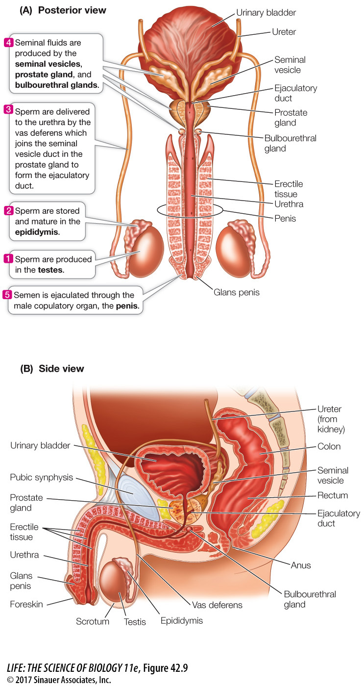
Semen contains sperm and a complex mixture of fluids and molecules (seminal fluid) that support the sperm and facilitate fertilization. Sperm make up less than 5 percent of the volume of the semen. Sperm are produced in the testes, the paired male gonads, and the seminal fluid is produced by accessory glands.
Figure 42.9 illustrates the human male reproductive organs. The testes of most mammals are located outside the body cavity in a pouch of skin called the scrotum. Why should the testes be located outside the body cavity? The optimal temperature for spermatogenesis in most mammals is slightly lower than the normal body temperature. The scrotum keeps the testes at this optimal temperature. Muscles in the scrotum contract in a cold environment, bringing the testes closer to the warmth of the body; in a hot environment the muscles relax, cooling the testes by suspending them farther from the body.
Question
Q: What problem might an enlarged prostate gland produce?
An enlarged prostate gland could squeeze the urethra shut and prevent or impair urination.
Activity 42.1 The Human Male Reproductive Tract
www.life11e.com/
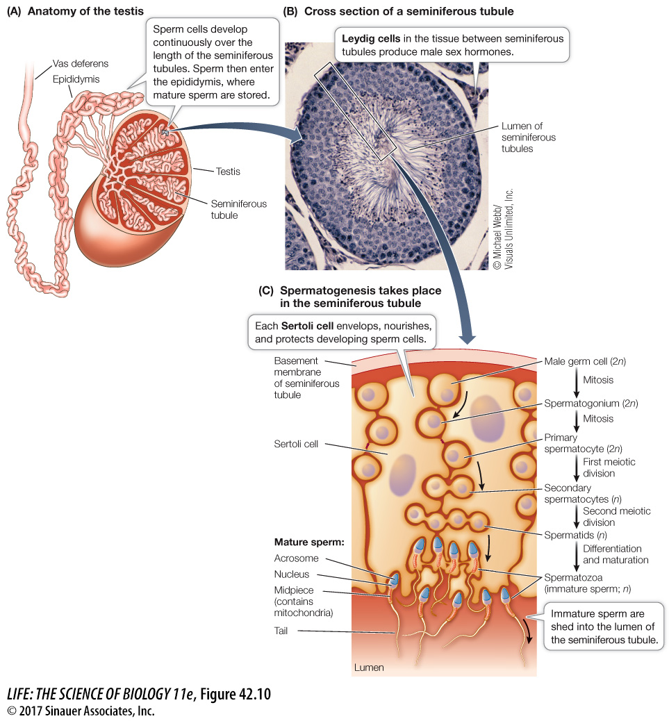
Spermatogenesis takes place within the seminiferous tubules tightly coiled in each testis (Figure 42.10A). Between the seminiferous tubules are clusters of Leydig cells that produce testosterone (Figure 42.10B). Spermatogonia reside in the outer regions of the seminiferous tubules, just under the basement membrane. Moving inward toward the lumen of the tubule are germ cells in successive stages of spermatogenesis (Figure 42.10C). The germ cells are intimately associated with Sertoli cells that provide nutrients for the developing sperm and secrete several things including hormones and growth factors that are necessary for spermatogenesis (which is why Sertoli cells are sometimes called “nurse cells”). Tight junctions (see Key Concept 6.2) between Sertoli cells create a blood–
Activity 42.2 Spermatogenesis
www.life11e.com/
When the second meiotic division is complete, each primary spermatocyte has produced four spermatids (see Figure 42.4A). The spermatids develop into spermatozoa as they migrate toward the lumen of the seminiferous tubule. The nucleus becomes compact, and the surrounding cytoplasm is lost. A flagellum—
From the seminiferous tubules, sperm move into the epididymis (see Figure 42.9A), where they mature, become motile, and are stored. The epididymis connects to the urethra via the vas deferens (plural vasa deferentia) and the ejaculatory duct. The urethra originates in the bladder, runs through the penis, and opens to the outside of the body at the tip of the penis. It serves as the common final duct for urine and semen.
902
The components of the semen other than sperm come from three accessory glands. About 60 percent of the volume of semen is secreted by the paired seminal vesicles that empty into the vas deferens just before it joins the urethra. Seminal fluid is thick because it contains mucus and fibrinogen, a protein also found in the blood, where it can polymerize to form blood clots. Seminal fluid also contains the monosaccharide fructose, an energy source for the sperm.
The prostate gland contributes about 30 percent of the volume of the semen. Prostate fluid is alkaline, so it neutralizes the acidity in the male and female reproductive tracts and makes these environments more hospitable to sperm. The prostate also secretes a clotting enzyme that causes fibrinogen from the seminal vesicles to convert the semen into a clotted, gelatinous mass, facilitating the semen’s propulsion into the upper regions of the female reproductive tract. Another prostate enzyme, fibrinolysin, then dissolves the clotted semen and liberates the sperm. Prostaglandins—
The bulbourethral glands produce a small volume of an alkaline, mucoid secretion that helps neutralize acidity in the urethra and lubricate it to facilitate the passage of semen during the climax of sexual intercourse.