key concept 42.4 Female Sex Organs Produce Eggs and Nurture Embryos
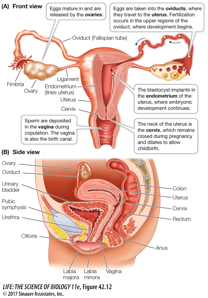
Figure 42.12 illustrates the human female reproductive system. Eggs are produced in and released from the ovaries located on each side of the lower abdominal cavity. When an egg leaves the ovary (ovulation), it enters the abdominal cavity, but it does not go far. The ovaries are close to the openings of the oviducts (also known as the Fallopian tubes). The openings are surrounded by undulating, fringed tissues called fimbria that sweep the egg into the oviduct. Cilia lining the oviduct propel the egg toward the uterus, a muscular, thick-
Activity 42.3 The Human Female Reproductive Tract
www.life11e.com/
focus your learning
The menstrual (uterine) cycle refers to the buildup and breakdown of the endometrial lining of the uterus.
If pregnancy occurs, human chorionic gonadotropin (hCG) stimulates the corpus luteum to continue producing estrogen and progesterone.
Mechanical and hormonal stimuli are involved in labor.
The ovarian and uterine cycles are coordinated and timed by the same hormones that initiate sexual maturation.
905
Externally, the vagina is enclosed by two sets of skin folds. The inner, more delicate folds are the labia minora; the outer, thicker folds are the labia majora. At the anterior tip of the labia minora is the clitoris, a small bulb of highly sensitive erectile tissue that has the same developmental origins as the male glans penis. Clitoral tissues, homologous to the erectile tissues of the penis extend deep under the labia minora and the walls of the vagina. The labia minora and the clitoral tissues become engorged with blood in response to sexual stimulation. Just as stimulation of the glans penis during sexual intercourse culminates in orgasm in the male, stimulation of the clitoral tissues results in orgasm in the female.
The external opening of an infant’s vagina is usually, but not always, partly covered by a thin membrane, the hymen. Eventually the hymen can be torn by vigorous physical activity or by first sexual intercourse; it can sometimes make first intercourse difficult or painful for the woman.
Sperm deposited in the vagina swim and are propelled by contractions of the female reproductive tract through the cervical opening, across the uterus, and most of the way up an oviduct, where fertilization takes place. Before meeting a sperm, the egg is still a secondary oocyte. Fertilization stimulates the egg to complete its second meiotic division. Following that division, the haploid nuclei of the sperm and the egg can fuse to produce a diploid zygote nucleus.
The *zygote undergoes its first few cell divisions to become a blastocyst while still in the oviduct. The blastocyst moves down the oviduct to the uterus, where it attaches itself to the epithelial lining of the uterus—
*connect the concepts The processes by which the zygote forms, develops into a blastocyst, and becomes implanted in the uterus are illustrated in Key Concept 44.2.
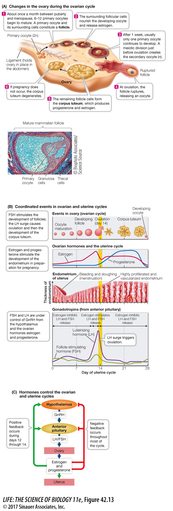
The maturation of eggs and ovulation are cyclical events in the ovaries. Changes in the uterus are also critical processes. These cycles in humans average about 28 days, but they can vary considerably in an individual and among different individuals. In anticipation of receiving a blastocyst, the endometrium thickens and develops lots of blood vessels. The endometrium regresses if a blastocyst does not arrive within a certain window of time. Thus female reproductive functions consist of two linked cycles: an ovarian cycle that produces eggs and hormones; and a uterine, or menstrual, cycle that prepares the endometrium for the arrival of a blastocyst (Focus: Key Figure 42.13). The two cycles must be synchronized so that a blastocyst arrives in the uterus at the optimal time to implant in the endometrium and continue its development.
906
focus: key figure
Question
Q: Hormonal contraceptive pills contain estrogen and progesterone, which portion of the ovarian cycle do they simulate and how do they prevent ovulation?
Hormonal contraceptive pills simulate the second half of the ovarian cycle, the luteal phase when estrogen and progesterone levels are high. These two hormones exert negative feedback that inhibits the release of gonadotropin, LH, and FSH which are essential for maturation of the egg and ovulation.
Animation 42.2 The Ovarian and Uterine Cycles
www.life11e.com/