Chapter Introduction
916
43
key concepts
43.1
Fertilization Activates Development
43.2
Mitosis Divides Up the Early Embryo
43.3
Gastrulation Generates Multiple Tissue Layers
43.4
Organs Develop from the Three Germ Layers
43.5
Extraembryonic Membranes Nurture Avian and Mammalian Embryos
Animal Development
investigating life
Go With the Flow
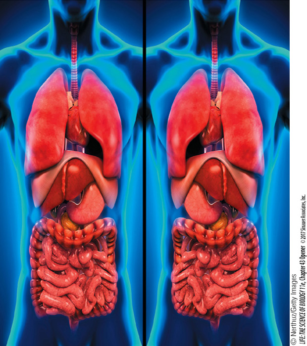
Place your hand over your heart. Point to your appendix, then your pancreas. Surely your hand went to the left side of your chest, then to the right side of your lower abdomen, and lastly to the left side of your upper abdomen. Vertebrates are bilaterally symmetrical, but our symmetry is not absolute. Some internal organs are oriented to the left and some to the right. In about 1 out of every 7,000 people, these orientations are reversed, a condition known as situs inversus (“location inverted”). The difference arises from events very early in the development of the embryo. Most people with situs inversus lead normal lives and are unaware of their condition until they have diagnostic imaging or surgery.
You will learn in this chapter that a mammalian embryo goes from a single layer of cells to the next stage, with two layers of cells, by forming a midline slit, and that cells migrate through that slit to form a second cell layer. Below where the inward movement of cells starts is an area called the node. Each cell at the node has a primary cilium. The primary cilia of some of these cells are nonmotile and probably serve sensory functions. The primary clila of other nodal cells are motile, and they beat in a clockwise motion that moves extracellular fluid across the surface of the node. That fluid movement is from right to left. Why?
Imagine that the movement of a cilium describes a clock face oriented so that 12 o’clock is closest to the embryo’s future head. The cilia protrude from the cell surfaces at an angle pointing posteriorly. Thus when the cilia rotate through the 6 o’clock position, they are closer to the cell surface and experience greater resistance than at the 12 o’clock position. As a result, the circular beating of cilia at this critical spot—
Why do we hypothesize that the information generated by beating nodal cilia is responsible for left–
How does the embryo know left from right?