The amount of yolk influences gastrulation
927
The eggs of reptiles, including birds, contain a mass of yolk, and the blastulas of these groups develop as a disc of cells on top of the yolk (see Figure 43.3B). We will use the chicken egg to show how gastrulation proceeds in a flat disc of cells rather than in a ball of cells.
Cleavage in the chick results in a flat, circular layer of cells called a blastodisc (Figure 43.11). Between the blastodisc and the yolk mass is a fluid-
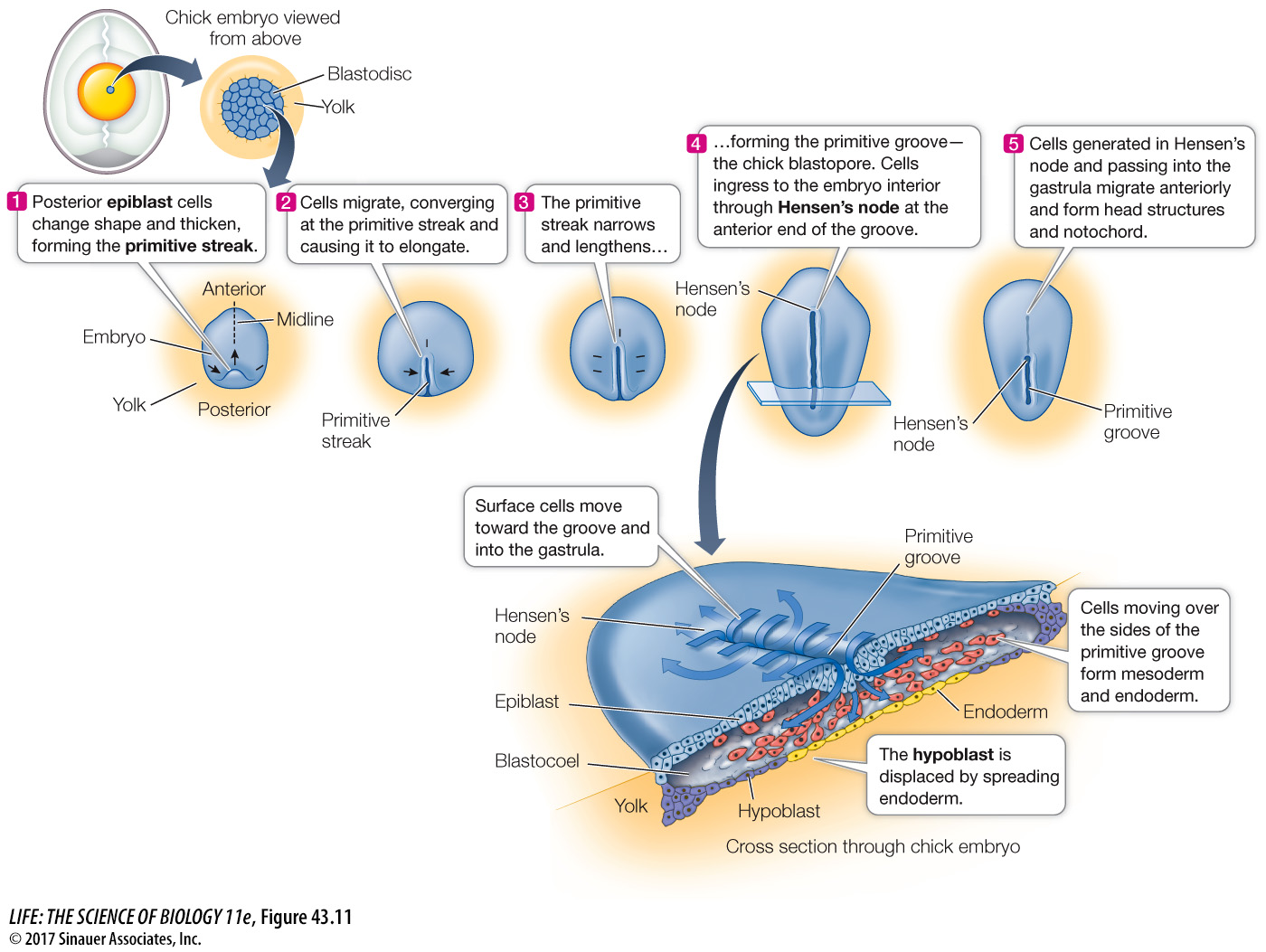
Gastrulation begins with a thickening in the posterior region of the epiblast, caused by the movement of cells toward the midline and then forward along the midline (see Figure 43.12). The result is a midline ridge called the primitive streak. A depression called the primitive groove forms along the length of the primitive streak. The primitive groove functions as the blastopore, and cells migrate through it into the blastocoel to become endoderm and mesoderm.
928
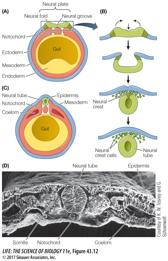
In the chick embryo, no archenteron forms, but the endoderm and mesoderm migrate forward to form the gut and other structures. At the anterior end of the primitive groove is a thickening called Hensen’s node, which in reptiles, birds, and mammals is the equivalent of the dorsal lip of the amphibian blastopore. Many signaling molecules that have been identified in the frog organizer are also expressed in Hensen’s node. Cells moving into the blastocoel and moving anteriorly from Hensen’s node become the notochord and organize the chick embryo in a manner similar to that of the frog embryo.