Cleavage produces a multicellular embryo
Cleavage is the cell divisions that transform the diploid zygote into a mass of undifferentiated cells that develop as the embryo. Zygote cytoplasm is not homogeneous, and these first cell divisions result in the differential distribution of nutrients and cytoplasmic determinants in the early embryo.
In most animals, cleavage proceeds with rapid DNA replication and mitosis but with no cell growth and little gene expression. The embryo becomes a solid ball of smaller and smaller cells. Eventually this ball forms a central fluid-
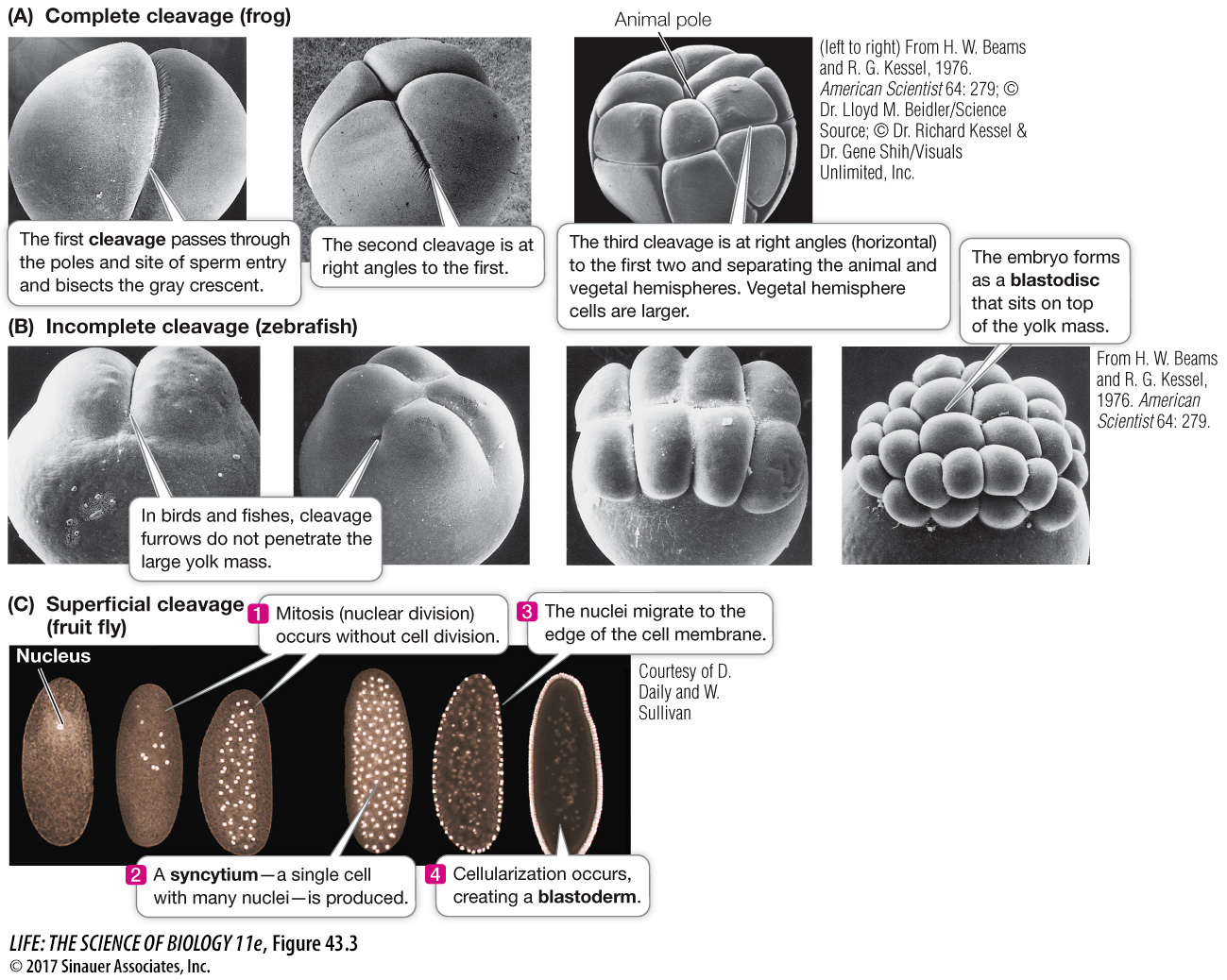
Complete cleavage occurs in eggs that have little yolk such as the eggs of mammals. Early cleavage furrows divide the egg completely. The amphibian egg undergoes complete cleavage, but vegetal pole yolk causes unequal cytoplasm division and animal hemisphere blastomeres are smaller than those in the vegetal hemisphere (Figure 43.3A).
Incomplete cleavage occurs in eggs with a lot of yolk and the cleavage furrows do not penetrate it completely. Discoidal cleavage is a type of incomplete cleavage common in fishes as well as in birds and other reptiles, in which the embryo forms as a disc of cells, or blastodisc, that sits on top of the dense yolk mass (Figure 43.3B).
Superficial cleavage is a variation of incomplete cleavage that occurs in insects such as the fruit fly (Drosophila). Early in development, cycles of mitosis occur without cell division, producing a syncytium—a single cell with many nuclei (Figure 43.3C). The nuclei eventually migrate to the periphery of the egg, after which the cell membrane of the egg grows inward, creating a blastoderm by partitioning the nuclei into individual cells surrounding a core of yolk.
920
The positions of the mitotic spindles during cleavage are not random but are defined by *cytoplasmic determinants produced from the maternal genome and stored in the egg. The orientation of the mitotic spindles can determine the planes of cleavage and the arrangement of the blastomeres.
*connect the concepts As described in Key Concept 19.2, cytoplasmic determinants are regulatory substances that are distributed unequally in the egg cytoplasm and that influence polarity and cell fate during embryogenesis.
In complete cleavage, if the mitotic spindles of successive cell divisions form parallel or perpendicular to the animal–