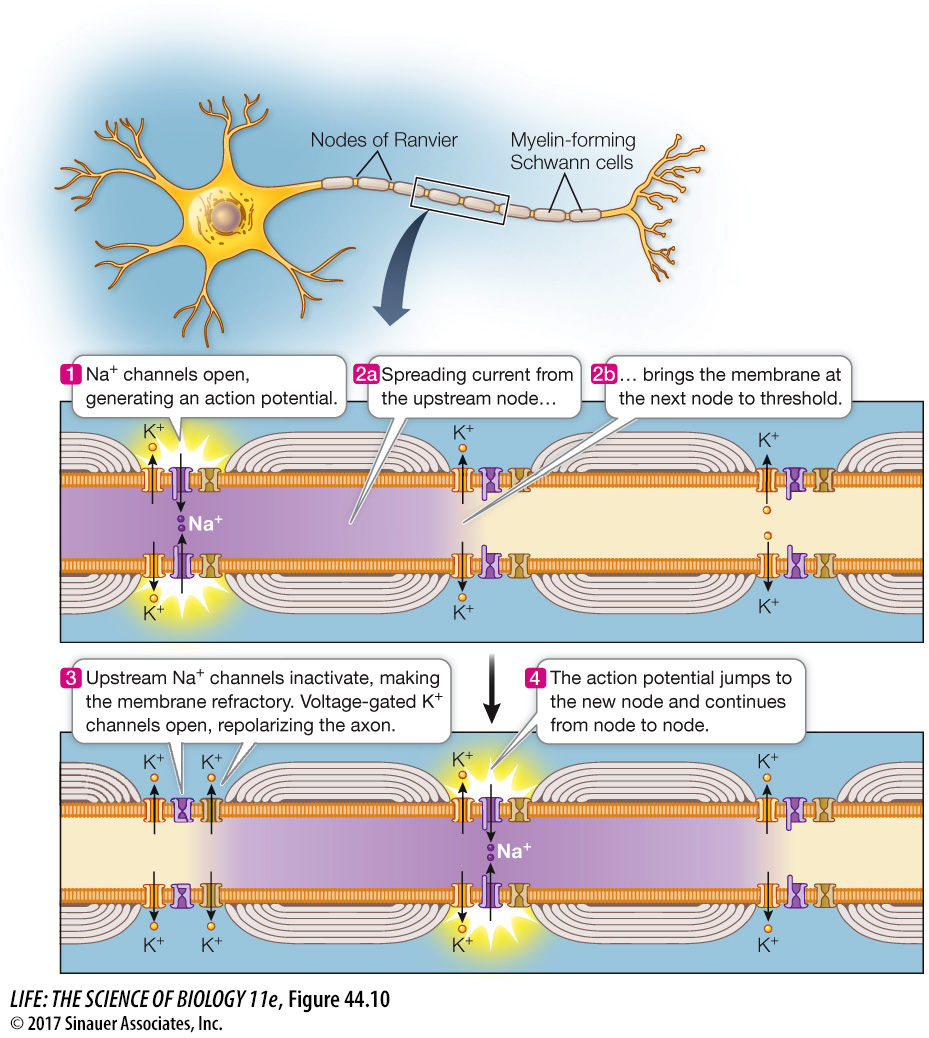
Action potentials are conducted along axons without loss of signal
An AP is conducted over long distances with no loss of signal. If we place two pairs of electrodes at two different locations along an axon, we can record an AP at those two locations as it travels along the axon (Figure 44.9A). The magnitude of the AP does not change between the two recording sites. This constancy is possible because an AP is an all-
948
An AP is all-
or- none because of the interaction between the voltage- gated Na+ channels and the membrane potential. If the membrane is depolarized slightly, some voltage- gated Na+ channels open. Some sodium ions cross the cell membrane and depolarize it even more, opening more voltage- gated Na+ channels, and so on, generating an AP. This positive feedback mechanism ensures that APs always rise to their maximum value. An AP is self-
regenerating because it spreads by local current flow to adjacent regions of the cell membrane. The resulting depolarization brings those neighboring areas of membrane to threshold. Therefore, when an AP occurs at one location on an axon, it stimulates the adjacent region of axon to generate an AP, and so on down the length of the axon. Although we say that the AP travels down the axon, what is really happening is that new APs are being created sequentially.
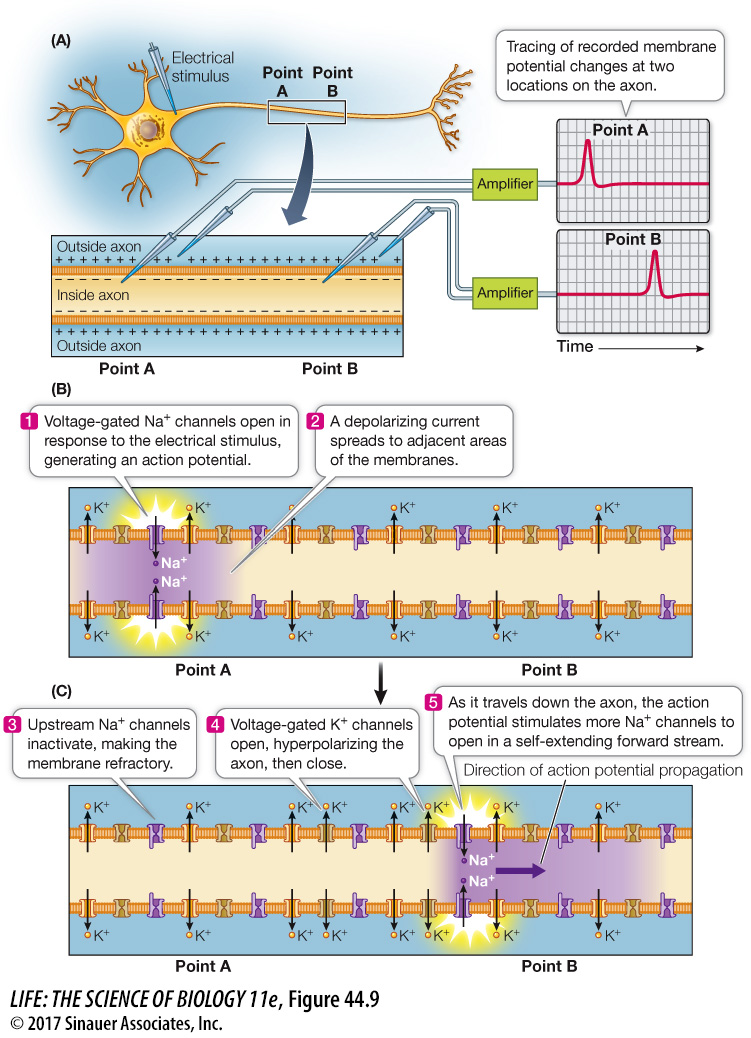
We can use an electrode to stimulate an axon, causing it to depolarize and to fire an AP that is then conducted along the axon. Figure 44.9B shows the changes in the ion channels in the membrane that are responsible for conducting the AP along the axon without a reduction in amplitude. Normally an AP is propagated in only one direction—
APs are not conducted at the same speed in all axons. They travel faster in large-