Auditory systems use hair cells to sense sound waves
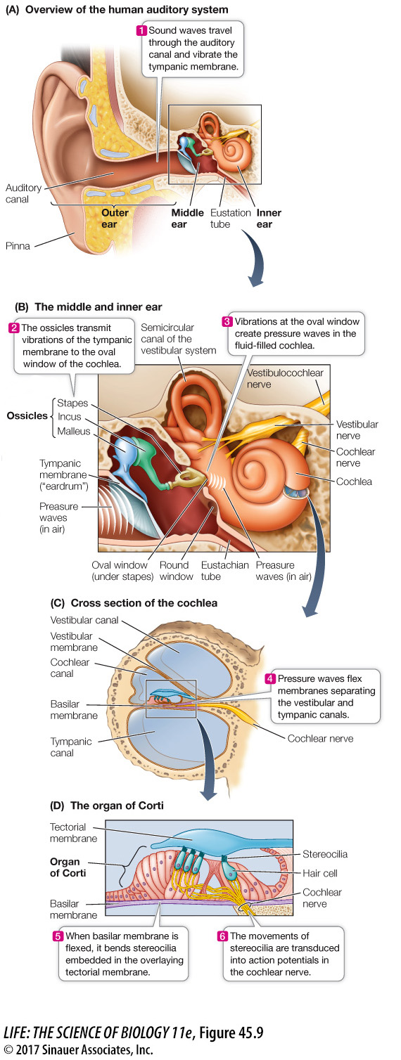
The stimuli that animals perceive as sounds are pressure waves. Auditory systems use mechanoreceptors to convert pressure waves into receptor potentials. Auditory systems include special structures that gather sound waves, direct them to the sensory organ, and amplify their effect on the mechanoreceptors. A good example of an auditory system is the human ear (Focus: Key Figure 45.9), which can be divided into three major areas: the outer, middle, and inner ear (see Figure 45.9A).
focus: key figure
Question
Q: How can the ears detect directionality of a sound source?
Several mechanisms can enable bilaterally placed ears to detect directionality of a sound: (1) The sound will be louder the closer the ear is to the source, and also the head creates a sound shadow for sounds coming from the side. (2) The sound will reach the closer ear sooner than the more distant ear. (3) If the sound is not coming from directly in front or in back, there will be a phase mismatch between the sound waves reaching the two ears.
Activity 45.1 Structures of the Human Ear
www.life11e.com/
OUTER EAR The outer ear consists of the pinnae (singular, pinna) and the auditory canal. The pinnae collect sound waves and direct them into the auditory canals; watch a dog change the orientation of its ears to focus on a particular sound to get the idea of the role pinnae play in hearing. Covering the end of the auditory canal is the tympanic membrane (commonly called the eardrum) which vibrates in response to pressure waves traveling down the canal, thus converting the pressure waves to physical forces in the middle ear.
969
MIDDLE EAR On the other side of the tympanic membrane is the middle ear, an air-
The middle ear contains the ossicles, three delicate bones individually named the malleus (“hammer”), incus (“anvil”), and stapes (“stirrup”) (see Figure 45.9B). The ossicles transmit the vibrations of the tympanic membrane to another flexible membrane, the oval window. The ossicles act as a lever (like a hammer pulling out a nail), translating a large movement of the tympanic membrane into a smaller movement of the oval window, but a movement of greater force. Because the oval window is much smaller than the tympanic membrane, the pressure the stapes transmits to the oval window is more than 20 times greater than the pressure exerted by a sound wave on the tympanic membrane.
Behind the oval window lies the fluid-
INNER EAR The inner ear is a bony structure consisting of two sets of canals. One is the organ of balance, the vestibular system, and the other is the organ of hearing, the cochlea. The cochlea (“snail” or “spiral shell”) is a long, tapered, coiled structure. A cross section of the cochlea reveals that it is composed of three parallel canals separated by two membranes, the vestibular membrane and the basilar membrane (see Figure 45.9C). Sitting on the basilar membrane is the organ of Corti, which transduces pressure waves into action potentials. The organ of Corti contains hair cells with stereocilia (see Figure 45.8). The tips of the hair cells are embedded in a gelatinous overhanging shelf called the tectorial membrane (see Figure 45.9D).
Stereocilia are not motile, but because their tips are attached to the more rigid tectorial membrane, stereocilia bend when the basilar membrane flexes. As shown in Figure 45.8, when the stereocilia are bent in one direction, the hair cell is depolarized by an inflow of K+ ions. This effect of opening K+ channels happens because the fluid in the cochlear canal has a higher K+ concentration than does the intracellular fluid of the hair cell. The response of the hair cell is a graded membrane potential. The hair cells do not fire action potentials, but the changes in their membrane potential alter the rate at which the hair cells release neurotransmitter onto the sensory neurons whose axons make up the auditory nerve and transmit action potentials to the brain.
The vestibular and tympanic canals are separate until they reach the distal end of the cochlea (the end farthest from the oval window), where they join; thus they form one continuous canal that turns back on itself (see Figure 45.10). Just as the oval window is a flexible membrane at the beginning of the vestibular canal, the round window is a flexible membrane at the end of the tympanic canal. When the oval window vibrates, the waves of fluid pressure create traveling waves of flexion in the basilar membrane.