Information flows through layers of neurons in the retina
977
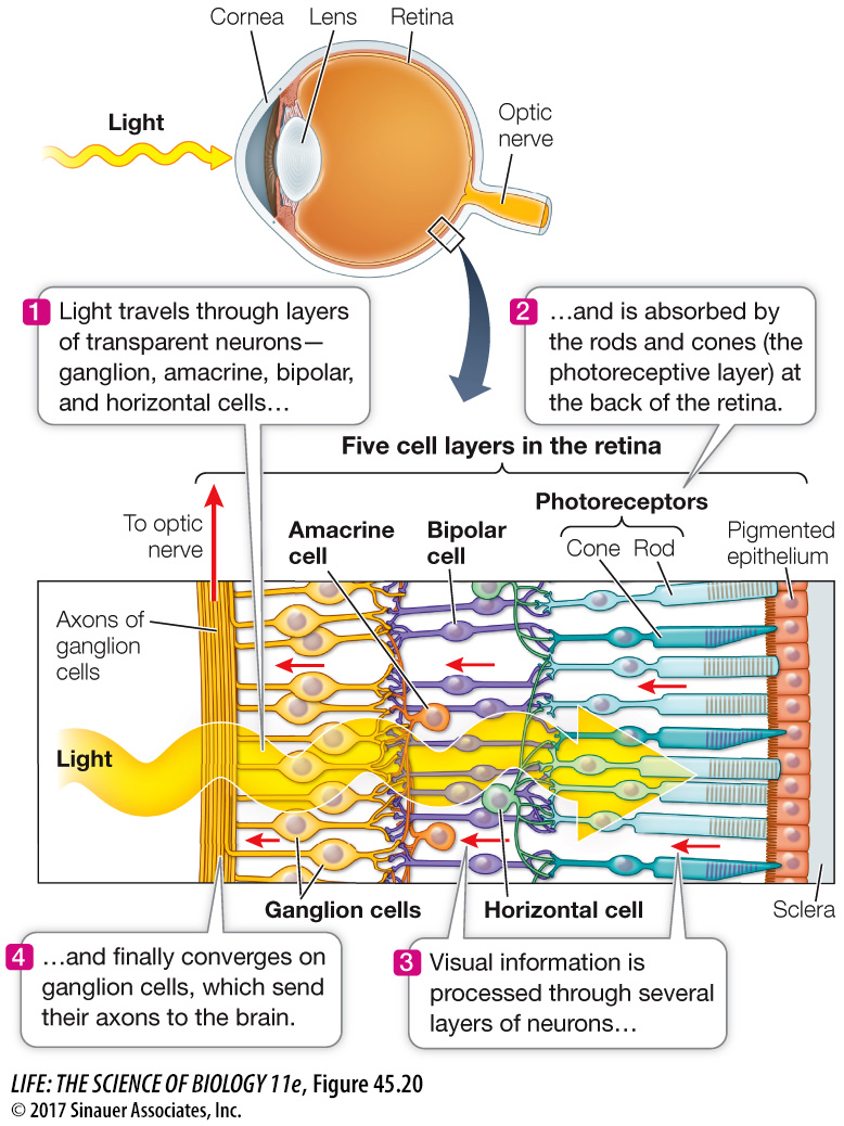
The human retina is organized into layers of neurons that receive visual information and process it before sending it to the brain (Figure 45.20). Closest to the lens (and thus to light input) is a layer of ganglion cells; a central layer contains three neuronal types, bipolar cells, horizontal cells, and amacrine cells; and at the “rear” of the retina lie the photoreceptors (rods and cones). The layers of cells between the photoreceptors and the ganglion cells process information about the visual field.
Media Clip 45.1 Into the Eye
www.life11e.com/
Activity 45.4 Structure of the Human Retina
www.life11e.com/
GANGLION AND BIPOLAR CELLS From our discussion of rod cells, you know that the photoreceptor cells at the back of the retina hyperpolarize in response to light and do not generate action potentials. The ganglion cells at the front of the retina do, however, fire action potentials. The axons of ganglion cells form the optic nerves that travel to the brain.
The ganglion cells are connected to the photoreceptors by bipolar cells. Changes in the membrane potential of rods and cones in response to light alter the rates at which the rods and cones release neurotransmitter at their synapses with the bipolar cells. In response to this neurotransmitter, the membrane potentials of the bipolar cells change, altering the rate at which they release neurotransmitter onto ganglion cells. The neurotransmitter release from the bipolar cells determines the rate at which ganglion cells fire action potentials. Thus the direct flow of information in the retina is from photoreceptor to bipolar cell to ganglion cell. The ganglion cells send the information to the brain via the optic nerves.
Each human eye contains about 1.2 million ganglion cells but more than 100 million rods and cones. Therefore there must be convergence of information as it passes from the photoreceptors to the ganglion cells. A given bipolar cell can receive input from multiple rods or multiple cones, but not from both. The relationship between photoreceptors, bipolar cells, and ganglion cells depends on their location on the retina. In the fovea, a ganglion cell may receive input from as few as five photoreceptors, but in the periphery of the retina, a ganglion cell may receive input from thousands of photoreceptors. Visual acuity is a reflection of these quantitative relationships.
The patch of photoreceptors that communicates with a ganglion cell forms a circular receptive field. When light falls on a receptive field, its ganglion cell can be either excited or inhibited. As mentioned above, each ganglion cell sends an axon to the brain in the optic nerves. Thus the information coming from the retina to the brain is about the pattern of patches of light and dark falling on the retina. We’ll return to a discussion of how the brain constructs the visual field from the input from receptive fields in Chapter 46.
HORIZONTAL AND AMACRINE CELLS The other two cell layers, the horizontal cells and the amacrine cells, consist of interneurons that communicate laterally across the retina. Horizontal cells form synapses with neighboring photoreceptors and bipolar cells. Thus light falling on one photoreceptor can influence the sensitivity of its neighbors to light. This lateral flow of information enables the retina to sharpen the perception of contrast between light and dark patterns. Amacrine cells form local interconnections between bipolar cells and ganglion cells. Some amacrine cells are highly sensitive to changing illumination or to motion. Others assist in adjusting the sensitivity of the eyes according to the overall level of light falling on the retina. When background light levels change, amacrine cell connections to the ganglion cells adjust the range of intensities to which they are sensitive. Thus even with large changes in background illumination such as going from outdoors into a building, the eyes are still sensitive to small, rapid changes in the pattern of light falling on the retina. The light reflected from this page can vary by nine orders of magnitude (1,000,000,000-