Pathways of the autonomic nervous system control involuntary physiological functions
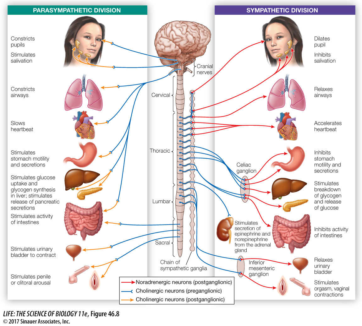
The autonomic nervous system, or ANS, includes CNS and PNS components and controls many involuntary functions, such as heart rate, blood flow, sweating, and digestive activities. Its control of diverse organs and tissues is crucial to homeostasis. The ANS has two divisions, sympathetic and parasympathetic, that work in opposition to each other in their effects on most organs (Figure 46.8). The sympathetic and parasympathetic divisions are easily distinguished by their anatomy, neurotransmitters, and actions.
Question
Q: Why does a person get an extremely dry mouth when speaking in public?
When an individual is stressed such as when speaking in public, the sympathetic nervous system is activated (fight-
The best-
Whether sympathetic or parasympathetic, every autonomic efferent pathway begins with a cholinergic neuron (that is, a neuron that uses acetylcholine as its neurotransmitter) that has its cell body in the brainstem or spinal cord. These cells are called preganglionic neurons because the second neuron in the pathway with which they synapse resides in a collection of neurons outside the CNS called a ganglion (plural ganglia). The second neuron is called a postganglionic neuron because its axon extends out from the ganglion. The axon of the postganglionic neuron synapses with cells in the target organs (see Figure 46.8).
The postganglionic neurons of the sympathetic division mostly use norepinephrine (also known as noradrenaline) as their neurotransmitter. In contrast, the postganglionic neurons of the parasympathetic division are mostly cholinergic. In organs that receive both sympathetic and parasympathetic input, the target cells respond in an opposite manner to norepinephrine and to acetylcholine. This happens, for example, in a region of the heart called the pacemaker, which initiates the heartbeat. Stimulating the sympathetic nerve to the heart or dripping norepinephrine onto pacemaker cells increases their firing rate and causes the heart to beat faster. In contrast, stimulating the parasympathetic nerve to the heart or dripping acetylcholine onto pacemaker cells decreases their firing rate and causes the heart to beat more slowly (see Figure 49.7).
990
The sympathetic and parasympathetic divisions of the ANS can also be distinguished by anatomy. The preganglionic neurons of the parasympathetic division come from the cranial nerves of the brainstem and the sacral (lowest) region of the spinal cord; those of the sympathetic division come from the thoracic and lumbar regions of the spinal cord (see Figure 46.8). Most of the ganglia of the sympathetic division are lined up and interconnected in two chains, one on either side of the spinal cord. The parasympathetic ganglia are close to the target organs and are not interconnected.
The ANS is an important link between the CNS and many physiological functions. Its control of diverse organs and tissues is crucial to homeostasis. Despite the complexity of the ANS, work by neurobiologists and physiologists over many decades has made it possible to understand its functions in terms of neuronal properties and circuits.