Three-
How do we perceive objects in three dimensions? The short answer is that our two eyes see overlapping, yet slightly different, visual fields—
992
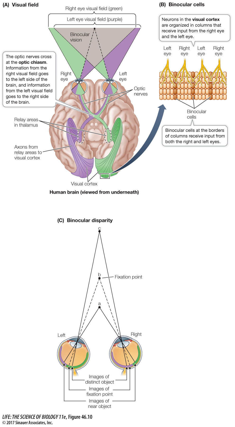
The story of how the brain integrates information from two eyes begins with the paths of the optic nerves. The two optic nerves run along the underside of the brain, join just under the hypothalamus, and then separate again (Focus: Key Figure 46.10). The place where they join is called the optic chiasm. Axons from the half of each retina closest to the nose cross in the optic chiasm and go to the opposite side of the brain. The axons from the outer half of each retina do not cross over at the optic chiasm; axons from the outer left retina go to the left side of the brain, and vice versa for axons from the outer right retina.
993
focus: key figure
Question
Q: What would be the consequences of an anterior–
The right and the left visual fields would be narrower and there would be no depth perception.
The functional consequence of the optic chiasm is that all of the visual information from the left side of your field of vision when you are looking straight ahead goes to the right side of your brain, and all of the visual information from the right side of your field of vision goes to the left side of your brain. However, there is a very large area of overlap between the visual fields of the two eyes. These relationships are shown in purple and green in Figure 46.10.
Because of the overlap between the visual fields of the two eyes, there can be neurons in the visual cortex that receive input from both the left and the right eye. These are called binocular cells, and they can detect disparity between where the image of an object falls on the two retinas. What is disparity? Hold your finger out in front of you and look at it closing one eye and then the other. Your finger appears to jump back and forth because its image falls on a different position on each retina. Repeat the exercise with a distant object. It doesn’t jump back and forth as much because there is less disparity in the positions of the image on the two retinas. Certain binocular cells respond optimally to a stimulus falling on both retinas with a particular disparity. Which set of binocular cells is stimulated depends on how far away the stimulus is.
When we look at something, we can detect its shape, color, depth, and movement. Where does all this information come together? Is there a single cell that fires only when a red sports car drives by? The answer to that is probably no. Visual experience comes from simultaneous activity in many different cells. In addition, most visual experiences are enhanced by information from the other senses and from memory, which helps explain why about 75 percent of the cerebral cortex is association cortex.