Bones develop from connective tissues
Bones are divided into two types on the basis of how they develop. Membranous bone forms on a scaffold of connective tissue membrane. Cartilage bone forms first as a cartilaginous structure resembling the future mature bone, then gradually hardens, or ossifies, to become bone. The outer bones of the skull are membranous bones; the bones of the limbs are cartilage bones.
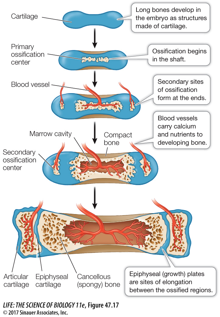
Cartilage bones can grow throughout the ossification process. The long bones of the legs and arms, for example, ossify first at the centers and later at each end (Figure 47.17). The cartilage between these ossification centers forms epiphyseal plates. Growth can continue until these areas of ossification join, eliminating the epiphyseal plates. The membranous bones forming the skull cap grow until their edges meet. The large soft spot on the top of a baby’s head, called a fontanel, is a point where the skull bones have not yet joined. There are multiple fontanels in the neonate where the skull bones eventually join.
1017
The structure of bone may be compact (solid and hard) or cancellous (having numerous internal cavities that make it appear spongy, although it is rigid). The architecture of a specific bone depends on its position and function, but most bones have both compact and cancellous regions. The shafts of the long bones of the limbs, for example, are cylinders of compact bone surrounding central cavities that contain fatty bone marrow called yellow bone marrow. The rigid, tubelike shaft of compact bone can withstand compression and bending forces. Architects and nature alike use hollow tubes as lightweight structural elements.
The ends of the long bones are cancellous (see Figure 47.17). Cancellous bone is lightweight because of its numerous cavities. These cavities contain the red bone marrow responsible for red blood cell formation as well as other cellular elements of the blood. Cancellous bone is also strong because its internal meshwork constitutes a support system. It can withstand considerable forces of compression.
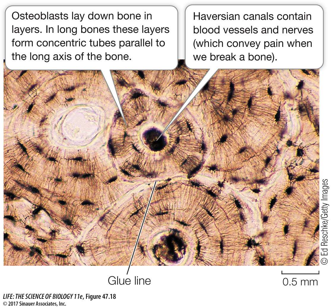
Most compact bone in mammals is called Haversian bone because it is composed of structural units called Haversian systems (Figure 47.18). Each Haversian system is a set of thin, concentric bony cylinders that surround a narrow canal, the Haversian canal, that contains blood vessels and nerves. Osteocytes are arranged in small spaces between the concentric circles of bone surrounding the canals. Adjacent Haversian systems are separated by boundaries called glue lines. Haversian bone is resistant to fracturing because cracks tend to stop at glue lines.