Hemoglobin’s affinity for O2 is variable
Various factors influence the O2-binding/dissociation properties of hemoglobin, thereby influencing O2 delivery to tissues. Three of these factors are the chemical composition of the hemoglobin, the blood pH, and the presence of 2,3-
HEMOGLOBIN COMPOSITION There is more than one type of hemoglobin because the chemical composition of the polypeptide chains that form the hemoglobin molecule varies. The normal hemoglobin of adult humans has two each of two kinds of polypeptide chains—
Before birth, the human fetus has a different form of hemoglobin, consisting of two α-globin and two γ-globin chains. The functional difference between fetal and adult hemoglobin is that fetal hemoglobin has a higher affinity for O2. Therefore the fetal hemoglobin–
HEMOGLOBIN AND pH The O2-binding properties of hemoglobin are also influenced by physiological conditions. The influence of pH on the function of hemoglobin is known as the Bohr effect. As blood passes through metabolically active tissue such as exercising muscle, it picks up acidic metabolites. As a result, blood pH falls. The excess H+ ions bind preferentially to deoxygenated hemoglobin and decrease its affinity for O2, and the O2-binding/dissociation curve of hemoglobin shifts to the right (see Figure 48.13). This shift means the hemoglobin will release more O2 in tissues where pH is low—
2,3-
When humans go to high altitudes, or when they cease being sedentary and begin to exercise, the level of 2,3-
1037
investigating life
Seals Are Champion Breath-
experiment
Original Paper: Castellini, M. A., G. L. Kooyman and P. J. Ponganis. 1992. Metabolic rates of freely diving Weddell seals: Correlations with oxygen stores, swim velocity, and diving duration. Journal of Experimental Biology 165: 181–
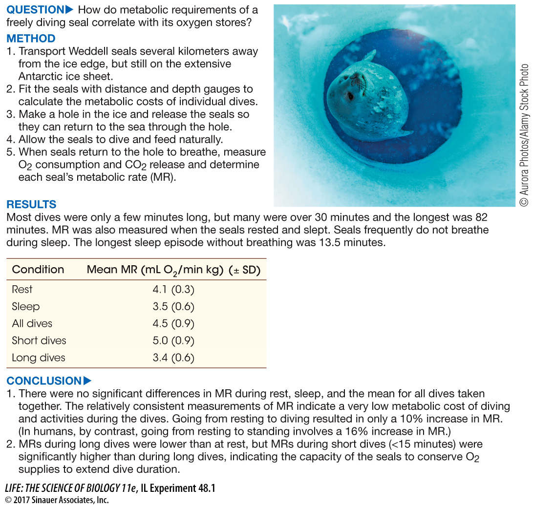
Weddell seals live near the edges of the ice in Antarctica where they feed by diving in the surrounding ocean. The ice-
work with the data
How do the seal’s oxygen stores compare with its oxygen demands? To calculate the oxygen reserves of a Weddell seal, we have to assess the amount of oxygen in its blood and in the myoglobin in its muscles at the beginning of the dive. The amount of oxygen in its lungs is irrelevant since seals exhale before diving, so there is little oxygen available in its alveoli during the dive.
The problem requires calculating the amount of O2 in the arterial and venous blood and myoglobin at the beginning of the dive that is available to support the seal’s MR.
Assume the following:
Average body mass: 355 kg
Blood volume: 14.8% of mass (7% in humans)
Hematocrit: 58% (human averages 42%)
Hemoglobin content of red blood cells: 23.7%
O2 holding capacity of hemoglobin: 1.34 mL/g
Blood: 34% is arterial, 95% saturated at beginning of dive, can desaturate to 20%
Available O2 in arterial blood is calculated as follows:
Total arterial blood = 355 kg × 0.148 × 0.34 = 17.9 kg
Total arterial hemoglobin = 17.9 kg × 0.58 × 0.24 = 2.49 kg
Maximum O2 content = 3.34 L
Arterial O2 available = 3.34 × (95% – 20%) = 2.5 L
QUESTIONS
Question 1
Calculate the available O2 in the venous blood. Assume venous blood is 66% of total blood volume, and it is 90% saturated at the beginning of the dive and can desaturate to 0%.
O2 available in venous blood:
Total venous blood = 355 kg × 0.148 × 0.66 = 34.7 kg
Total venous hemoglobin = 34.7 kg × 0.58 × 0.24 = 4.8 kg
Total venous O2 available = 4.8 kg × 1.34 L/kg × 0 .9 = 5.8 L
Question 2
Calculate the available O2 in myoglobin. Assume 33% of body mass is muscle, myoglobin content of seal muscle is 45 g/kg (20–
O2 available in myoglobin:
Total amount of myoglobin: 355 kg × 0.33 × 44g/kg = 5.2 kg
O2 bound to myoglobin: 5.2 kg × 1.34 L/kg = 7 L
Question 3
What are the total O2 reserves at the beginning of the dive?
Total O2 reserves at beginning of dive:
2.5 L + 5.8 L + 7.0 L = 15.3 L
Question 4
Given the MR values found in the experiment, what do you predict would be the maximum duration of no breathing during sleep?
Maximum sleep episode = total O2 reserves divided by the diving metabolic rate. So, 15.3 L divided by the rate of O2 consumption which for a long dive would be 0.0035 L/kg min × 355 kg. So, 15.3L/ 1.24 L/min = 12.3 min, which corresponds well with the observed maximum sleep episode of 13.5 min.
Question 5
Using the table of MR values for all dives in the results portion of the experiment, predict the maximum duration of a Weddell seal dive.
Assuming the overall dive metabolic rate of 4.5 mL O2/min kg, the maximum dive time would be 15.31/(0.0045 mL O2/min × 355 kg) = 10 min, which is much less than the observed dive times.
A similar work with the data exercise may be assigned in LaunchPad.