The ECG records the electrical activity of the heart
1056
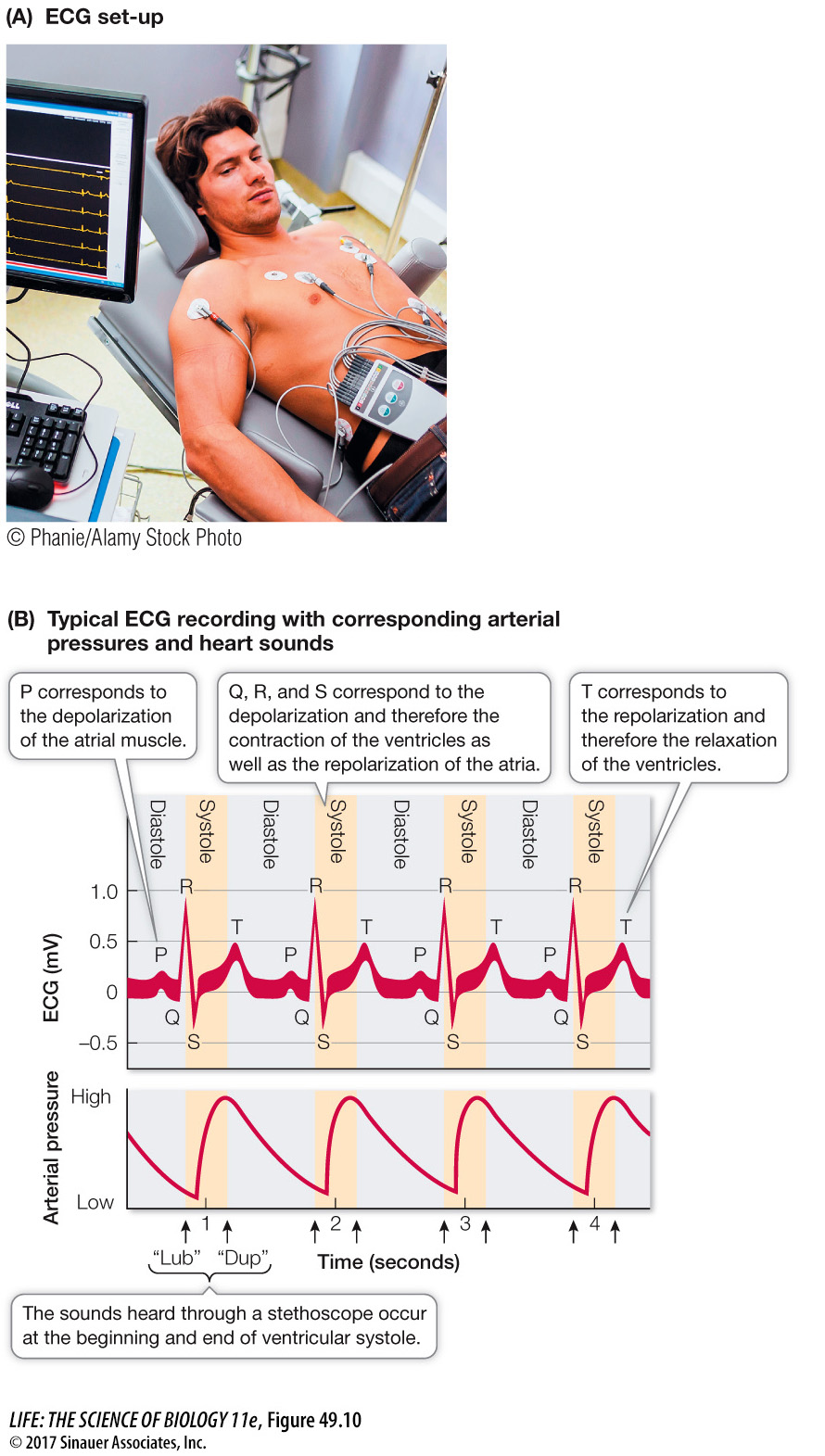
Electrical events in the cardiac muscle during the cardiac cycle are recorded by electrodes placed on the surface of the body. Such a recording is an electrocardiogram, or ECG, or EKG reflecting the Greek spelling (kardia). The ECG is an important tool for diagnosing heart problems (Figure 49.10A).
Question
Q: Abnormal ECG patterns are characteristic of HCM hearts. If there were an occasional or a long-
An occasional block of the AV node could result in a lengthening of the interval between the P and R waves (the PR interval) on the ECG and possibly a missed beat. A long-
The action potentials that sweep through the muscles of the atria and ventricles before they contract are massive, localized electrical events causing electric currents to flow throughout the body. Electrodes placed at different locations on the skin detect those currents at different times and register a voltage difference between them. The appearance of the ECG depends on the placement of the electrodes. Electrodes placed on the right wrist and left ankle produced the normal ECG shown in Figure 49.10B. The wave patterns of the ECG are designated P, Q, R, S, and T, each letter representing a particular event in the cardiac muscle, as shown in the figure.