Kidneys produce urine and the bladder stores it
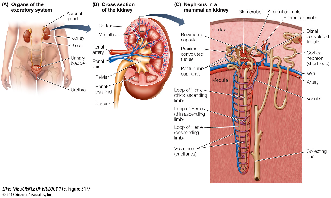
Mammalian excretory systems are similar, so we will use that of humans as our example. Humans have two kidneys at the back of the upper region of the abdominal cavity (Figure 51.9A). Each kidney filters blood, processes the filtrate into urine, and releases that urine into a duct called the ureter. The ureter of each kidney leads to the urinary bladder, where the urine is stored until it is excreted through the urethra, a short tube that opens to the outside of the body.
Question
Q: How do you think the osmolarity of the blood in the vasa recta compares with the osmolarity of the interstitial fluid at different levels of the medulla?
The blood flowing down the vasa recta always has a slightly lower osmolarity than the interstitial fluid at the same level because it is flowing and the equilibration is not instantaneous. The opposite is true for the ascending limb of the vasa recta, where the blood always has a slightly higher osmolarity than the surrounding interstitial fluid. As a result, the blood in the ascending limb absorbs water osmotically and carries some excess salt and water out of the renal medulla.
Activity 51.3 The Human Excretory System
www.life11e.com/
Two sphincter muscles surrounding the base of the urethra control urination. One of these sphincters is a smooth muscle and is controlled by the autonomic nervous system. As the bladder fills, stretch receptors in the walls of the bladder trigger a spinal reflex that relaxes this sphincter. This reflex is the only control of urination in infants, hence their frequent “accidents.” The other sphincter is a skeletal muscle and is controlled by the voluntary nervous system. When the bladder is very full, only deliberate conscious effort prevents urination. Toilet training of children teaches them to control this sphincter.