Chapter 43
RECAP 43.1
The sperm contributes the centriole to the embryo, and the centriole is the origin of the microtubules of the primary cilia, which serve signaling functions.
The unfertilized frog egg has an animal (upper) and a vegetal (lower) hemisphere. The outer cytoplasm of the animal hemisphere is pigmented, but the vegetal hemisphere cytoplasm is not. The sperm binds to the animal hemisphere and stimulates rotation of the outer (cortical) cytoplasm toward the site of sperm entry. This rotation creates a band of more lightly pigmented cytoplasm opposite the site of sperm entry.
In the unfertilized frog egg, β-catenin and its degrading enzyme GSK-
3 are homogeneously distributed. With sperm entry and cortical rearrangement, vesicles containing a GSK- inhibiting protein are translocated from the vegetal pole to the site opposite sperm entry. That protein regionally prevents the degradation of β-catenin on the dorsal side of the blastula.
A-
RECAP 43.2
Complete cleavage occurs in eggs that have little yolk. In these eggs the early cleavage furrows can divide the egg completely. In eggs with a lot of yolk, the cleavage furrows cannot penetrate completely and the blastula forms as a disc of cells that sits on top of the yolk mass.
Cleavage in mammals is very slow, allowing enough time for gene expression to occur. In sea urchins and frogs, cleavage occurs very rapidly, precluding gene expression between cell divisions.
Mammals have regulative development, meaning that each cell of the blastula is not irreversibly determined, so if the blastula is divided in half, identical twins can develop. Since development is mosaic in most invertebrates, if the blastula is divided, each part will be lacking certain elements and will not be able to complete development.
Germ cells are determined at the time of early cell divisions of the embryo that take place during formation of the blastula. These presumptive germ cells will not differentiate until the gonads develop and they migrate into those gonads. The origin of skin cells is ectoderm that is determined during a later developmental stage—
the gastrula— and these ectodermal cells differentiate rapidly to form the skin of the embryo.
RECAP 43.3
Gastrulation in both the sea urchin and the frog proceeds with an involution forming a blastopore. In the sea urchin, the cells involuting are similarly determined, but in the frog, tissue interactions differ according to location around the blastopore, and the involuting cells become differentially determined.
The criterion of necessity means that a component of a process must be present for that process to occur. Spemann’s experiment, constricting the fertilized egg so that the gray crescent was only in one daughter cell or in both daughter cells, showed that the gray crescent was necessary for embryo development. The criterion of sufficiency means that a component of a process can alone cause the process to occur. In the Spemann–
Mangold experiments, sufficiency was demonstrated by showing that when the dorsal lip of the blastopore was transplanted to another location on the blastula, it stimulated another axis of development. The first involuting cells of the primary organizer express the transcription factor Goosecoid, and as those cells move anteriorly, Goosecoid suppresses the expression of certain transcription factors in neighboring tissues that have to be suppressed so that organs appropriate for the head region can develop. Cells that leave the dorsal lip of the blastopore later express different transcription factors that also have suppressive action on expression of other transcription factors in neighboring tissues, enabling the induction of region-
appropriate organs. Gastrulation in reptiles and amphibians differs because the blastula of amphibians is a sphere, but the equivalent stage of reptilian development is a flat sheet of cells, the blastodisc. In amphibians, the cell movements of gastrulation take place through a hole in the blastula (blastopore), while in reptilian blastodiscs, a longitudinal slit (primitive streak) forms and the cell movements of gastrulation take place through that slit. In amphibians, the ingress of cells through the blastopore creates the archenteron (primitive gut). No archenteron forms in the reptilian embryo, but the endodermal and mesodermal cells that pass through the primitive streak migrate forward to become gut and other structures.
RECAP 43.4
The ectoderm over the notochord thickens on either side of the midline to form ridges aligned on an anterior−posterior axis. These ridges grow until they meet on the midline, forming the neural tube. The most lateral cells of these ridges break free to form neural crest cells, while still more lateral ectoderm comes together and merges to cover the neural tube.
A somite is a segmental and bilateral block of mesodermal cells that gives rise to vertebrae, ribs, trunk muscles, and limbs.
Vertebrates have groups of Hox genes arranged in linear order on multiple chromosomes. These genes are expressed along the anterior−posterior axis in the same sequence as they occur on the chromosomes. As a result, each anterior−posterior region of the embryo receives different combinations of Hox gene products in different amounts, producing a large combinatorial mix of signaling molecules.
RECAP 43.5
The yolk sac and allantois grow out from the hypoblast and consist of endoderm and mesoderm. The chorion and amnion develop from ectoderm and mesoderm.
The blastocyst divides into a trophoblast group and a hypoblast group of cells. The trophoblast interacts with the uterine lining to embed the embryo into the endometrium and to send out projections to make contact with maternal blood vessels. Cells of the hypoblast grow out to form what in birds would be the yolk sac, but there is no yolk. These hypoblast cells and the trophoblast cells form the placenta.
The first trimester is a time of great sensitivity of the embryo to environmental risks because this is the time when many signaling cascades are setting up subsequent processes of determination, differentiation, and development. Therefore the earlier any necessary lifestyle or environmental changes are made, the better.
WORK WITH THE DATA, P. 929
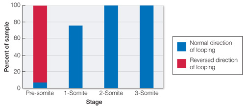
The experiment did not include left flow because we know left flow is the normal stimulus and that it is only disrupted by a fast right flow. This experiment was aimed at determining the sensitive period for the ability of that disruptive stimulus to alter the normal developmental pattern.
The data from embryos of different developmental stages support the conclusion that there is a sensitive phase of development where the left–
right symmetry of the embryo is determined, and that it is between the presomite and 1- somite stages. The results on 1-
, 2- , and 3- somite wild- type embryos exposed to fast right flow show that the left– right asymmetry was determined at an earlier stage and that the embryo is no longer sensitive to direction of flow over the node.
FIGURE QUESTIONS
Figure 43.7 Gastrulation involves the involution of vegetal pole cells over the lip of the blastopore to form the archenteron thus forming the lining of the gut.
Figure 43.8 Epiboly spreads the animal pole cells over the entire embryo and results in the formation of ectoderm, the cells that form the skin and structures associated with the skin.
APPLY WHAT YOU’VE LEARNED
No, the symptoms are not consistent with vitamin A deficiency. The baby’s defects are in mesenchyme tissues. A deficiency of vitamin A would most likely lead to defects of tissues derived from the ectoderm. In fact, the defects exhibited are consistent with an excess of vitamin A.
The ectoderm and the mesoderm express different genes. This results in mesoderm-
derived cells being sensitive to excesses of vitamin A and ectoderm- derived cells being sensitive to too little vitamin A. Individuals with this variant may be expected to be less susceptible to valproic acid. Because the variant enhances progression through G1 phase, the effect of valproic acid impeding this progression should be mitigated.
Even for women who have not been exposed to valproic acid, supplements of folic acid have been shown to reduce the frequency of neural tube defects. Given that valproic acid exposure increases risk for these defects, additional supplements of folic acid may counteract the effect of valproic acid. The extent to which additional folic acid could help would depend in part on whether and how these chemicals are involved in the same developmental processes. Valproic acid may have effects early in pregnancy—
perhaps even before the woman knows she is pregnant— because neural tube formation is affected by processes that occur early in pregnancy. Because deficiencies of folic acid increase the risk of neural tube defects, the efficiency of the receptor should affect this risk. Individuals who have receptor proteins that are more efficient in bringing folic acid to the cell should have greater cellular availability of folic acid and thus be at lower risk for neural tube defects compared with individuals who have less efficient receptor proteins.