Chapter 5
RECAP 5.1
Life is continuous: the cell theory explains that this comes from cell continuity. Life is complex chemically: the cell theory states that this chemistry occurs within cells. Life evolves and changes over time: the cell theory states that evolution occurs in cells as they change genetically and pass on new characteristics to the next generation of cells.
The membranes that enclose cells and organelles create compartments. The cell membrane allows the chemical reactions of life to occur away from the surrounding environment; in particular, the cell membrane permits the internal environment to have a distinct composition and concentration. Organelles’ membranes permit specialized functions and keep harmful substances away from the rest of the cell (cytoplasm).
RECAP 5.2
The cell wall is composed of a large complex of polysaccharides and peptides. It supports the cell and gives it shape. The capsule is a slimy polysaccharide layer that encloses the cell walls of some prokaryotes and keep cells from drying out. It may be used to attach to other cells. The flagellum is composed of strands of a large protein called flagellin and is attached to the cell wall and membrane by a motor complex. It is used for cell movement.
The nucleoid consists of DNA and proteins. Cytoplasm contains all the other small and large molecules for life, dissolved or suspended in water.
A-
RECAP 5.3
Prokaryotic cells are smaller than eukaryotic cells and lack organelles.
(a) Four membranes: two in the chloroplast and two in the mitochondrion. (b) Two membranes: the lysosomal membrane and the cell membrane (via vesicle; the molecules do not themselves cross any membranes). (c) No membranes: ribosomes do not have membranes. However, if the ribosomes were associated with the endoplasmic reticulum (ER), the answer would be two membranes: one into the ER and another out of the ER.
Microtubules line the long axons of nerve cells, where they act as tracks for vesicles that carry substances down the neuron. Without microtubules, the contents of these vesicles cannot be delivered to their destination, which can result in nerve problems. Depolymerization of microtubules can interfere with cell divisions because microtubules are a key part of the mitotic spindle that is used to move chromosomes during cell division.
For a lysosomal enzyme, the pathway would be ribosome → interior of ER → Golgi apparatus → Golgi vesicles → lysosome.
For an extracellular protein (animal cell), the pathway would be ribosome → interior of ER → Golgi apparatus → Golgi vesicles → cell membrane → extracellular region.
RECAP 5.4
Initially, there is a digestion of molecules that attach the cell membrane to the extracellular matrix. Then enzymes are produced and secreted from the cell that digests the macromolecules of the extracellular matrix. The cell migrates by microfilaments.
A cell wall contains cellulose fibers which provide strong support yet allow a plant to have some flexibility to bend. The cell wall structure provides an extra barrier that helps protect the cell from infection, yet cell walls in some cells have pores that allow the flow of materials in and out of the cell.
Chemical analysis could identify a plant cell by the presence of cellulose in the extracellular matrix and an animal cell by the presence of collagen in the extracellular matrix. These two compounds are distinct to the two different types of cells.
RECAP 5.5
The cell membrane could fold inward and then pinch off, forming vesicles for the Golgi apparatus and flat sheets for the ER.
The endosymbiotic theory states that one cell engulfed another cell. Over time the engulfed cell lost some DNA and the ability to perform independently of the host cell. Thus the engulfed cell became dependent on the host cell for its survival.
WORK WITH THE DATA, P. 99
The tannins appear in the chloroplast thylakoids and then are transferred to the vacuole by vesicles that enclose the tannins.
The staining for chlorophyll and tannins is in the same place. Since chlorophyll is in the chloroplast, this is consistent with a chloroplast origin for tannins, as described in the answer to Question 1.
Staining and chemical analysis both showed tannins, as well as cholorophyll, in the lower fraction of organelles, so this is probably a chloroplast fraction that contains tannins.
WORK WITH THE DATA, P. 103
The reasoning behind these experiments was as follows: if microfilaments are essential for cell movement, movement should not occur in the presence of cytochalasin B; if microtubules are essential for cell movement, movement should not occur in colchicine; if cell movement requires the synthesis of new proteins, movement should not occur in cycloheximide; and if cell movement requires energy, movement should not occur in dinitrophenol. The latter three experiments were important controls to rule out involvement of the three processes involved. The cycloheximide + cytochalasin B and the dinitrophenol + cytochalasin B controls demonstrated that the drugs had independent effects on the cells.
The experiment with cytochalasin B implicated microfilaments in cell movement. The experiment with colchicine ruled out microtubules. The experiments with cycloheximide showed that new protein synthesis is not necessary for cell movement. The experiments with dinitrophenol showed that cell movement does not require new inputs of energy.
FIGURE QUESTIONS
Figure 5.9 Assembly of the ER from the nuclear envelope; transport between Golgi apparatus cisternae; transport from ER to Golgi apparatus; endocytosis; exocytosis; transport from Golgi apparatus to cell membrane
Figure 5.11 Cells with high energy requirements, such as muscle cells, would have a lot of mitochondria.
Figure 5.18 Nexin links cause cilia and flagella to bend when microtubule doublets try to slide past one another. Absence of nexin would result in reduced flagella and ciliary function. This is called immotile cilia syndrome.
APPLY WHAT YOU’VE LEARNED
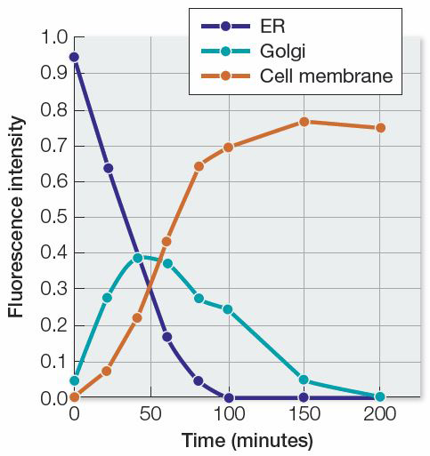
The results show that the protein begins its path in the endoplasmic reticulum, passes through the Golgi apparatus, and ends up in the cell membrane.
An inhibitor of protein synthesis would have prevented the fusion protein from being synthesized. Very little fluorescence (if any) would have been observed, and no fluorescence would have been observed moving into the Golgi apparatus or cell membrane.
The researchers would not have learned anything new because the cell fractionation method would have provided the same information about changes in subcellular locations of fluorescence over time. However, the results should have confirmed what was found using microscopy, which can provide additional weight of evidence for making claims about the path of the protein.
The researchers would have to use two different fluorescent tags that glow with different colors (e.g., green and red) so they could be distinguished from each other. Then the equipment used to collect data could be tuned to each color, and data collected at each color simultaneously.
As long as the two fluorescent tags on the different proteins can be distinguished from each other, they can be followed along different paths in the cell. The peroxidase would be expected to move from the ER to the Golgi apparatus to the peroxisome, while the secretory protein would be expected to move from the ER to the Golgi apparatus to the extracellular fluid. The two proteins would be found in the same compartments only in the ER and Golgi apparatus and would part ways as they entered different final destinations.
| Peroxidase | Secretory Protein | |||||||
|---|---|---|---|---|---|---|---|---|
| Time (min) |
ER | Golgi apparatus |
Peroxisome | Extracellular fluid |
ER | Golgi apparatus |
Peroxisome | Extracellular fluid |
| 0 | 1.0 | 0.0 | 0.0 | 0.0 | 1.0 | 0.0 | 0.0 | 0.0 |
| 10 | 0.6 | 0.3 | 0.1 | 0.0 | 0.6 | 0.3 | 0.0 | 0.1 |
| 20 | 0.4 | 0.4 | 0.2 | 0.0 | 0.4 | 0.4 | 0.0 | 0.2 |
| 50 | 0.2 | 0.3 | 0.5 | 0.0 | 0.2 | 0.3 | 0.0 | 0.5 |
| 100 | 0.1 | 0.2 | 0.7 | 0.0 | 0.1 | 0.2 | 0.0 | 0.7 |
| 200 | 0.0 | 0.0 | 1.0 | 0.0 | 0.0 | 0.0 | 0.0 | 1.0 |