Signal Amplification Occurs in the cAMP-PKA Pathway
We’ve seen that receptors such as the β-adrenergic receptor are low-abundance proteins, typically present in only a few hundred or thousand copies per cell. Yet the cellular responses induced by a hormone such as epinephrine can require production of large numbers of cAMP and activated enzyme molecules per cell. As an example, following activation of Gαs-coupled receptors, the intracellular concentration of cAMP rises to about 10−6 M. In a typical cell, that is roughly a cube about 15 µm on a side; this concentration is equivalent to 2 million molecules of cAMP per cell. Thus substantial amplification of the signal is necessary if it is to induce a significant cellular response. We have already seen how signal amplification occurs following photon absorption in rod cells. In the case of G protein–coupled hormone receptors, signal amplification is possible in part because both receptors and G proteins can diffuse rapidly in the plasma membrane. A single epinephrine-GPCR complex causes conversion of up to a hundred inactive Gαs molecules to the active form before epinephrine dissociates from the receptor. Each active Gαs·GTP, in turn, activates a single adenylyl cyclase molecule, which then catalyzes the synthesis of many cAMP molecules during the time Gαs·GTP is bound to it.
The amplification that occurs in such a signal transduction cascade depends on the number of steps in it and on the relative concentrations of the various components. In the epinephrine-induced cascade shown in Figure 15-7 and Figure 15-29, for example, blood levels of epinephrine as low as 10−10 M can stimulate liver glycogenolysis and release of glucose. An epinephrine stimulus of this magnitude generates an intracellular cAMP concentration of 10−6 M, an amplification of 104-fold. Because three more catalytic steps precede the release of glucose, another 104 amplification can occur, resulting in a 108 amplification of the epinephrine signal. In striated muscle, the amplification is less dramatic because the concentrations of the three successive enzymes in the glycogenolytic cascade—PKA, GPK, and GP—are in a 1:10:240 ratio (a potential 240-fold maximal amplification).
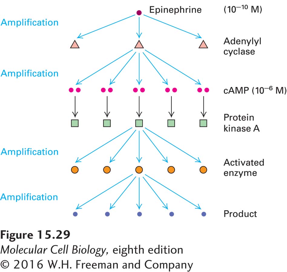
FIGURE 15-29 Amplification of an extracellular signal by a signal transduction pathway involving cAMP and PKA. The specific example here is the pathway depicted in Figure 15-7. Binding of a single epinephrine molecule to one G protein–coupled receptor induces activation of several molecules of adenylyl cyclase, the enzyme that catalyzes the synthesis of cyclic AMP, and each of these enzyme molecules synthesizes a large number of cAMP molecules, the first level of amplification. Two molecules of cAMP activate one PKA, but each activated PKA phosphorylates and activates multiple target proteins. This second level of amplification may involve several sequential reactions in which the product of one reaction activates the enzyme catalyzing the next reaction. The more steps in such a cascade, the greater the signal amplification possible.