Migrating Cells Are Steered by Chemotactic Molecules
Under certain conditions, extracellular chemical cues guide the locomotion of a cell in a particular direction. In some cases, the movement is guided by insoluble molecules in the underlying substratum, as in the wounded-cell monolayer assay described above. In other cases, the cell senses soluble molecules and follows them, along a concentration gradient, to their source—a process known as chemotaxis. For example, leukocytes are guided toward an infection by a tripeptide that is secreted by many bacterial cells (Figure 17-46). In another example, during the development of skeletal muscle, a secreted protein signal called scatter factor guides the migration of myoblasts to the proper locations in limb buds. One of the best-studied examples of chemotaxis is the migration of Dictyostelium amoebae during their starvation response. When these soil amoebae are stressed, they begin to secrete cAMP, which is an extracellular chemotactic agent in this organism. Other Dictyostelium cells move up the cAMP concentration gradient toward its source (see Figure 17-46). Thus the amoebae move toward one another, aggregate into a migratory slug, and then differentiate into a fruiting body in which starvation-resistant spores are formed.
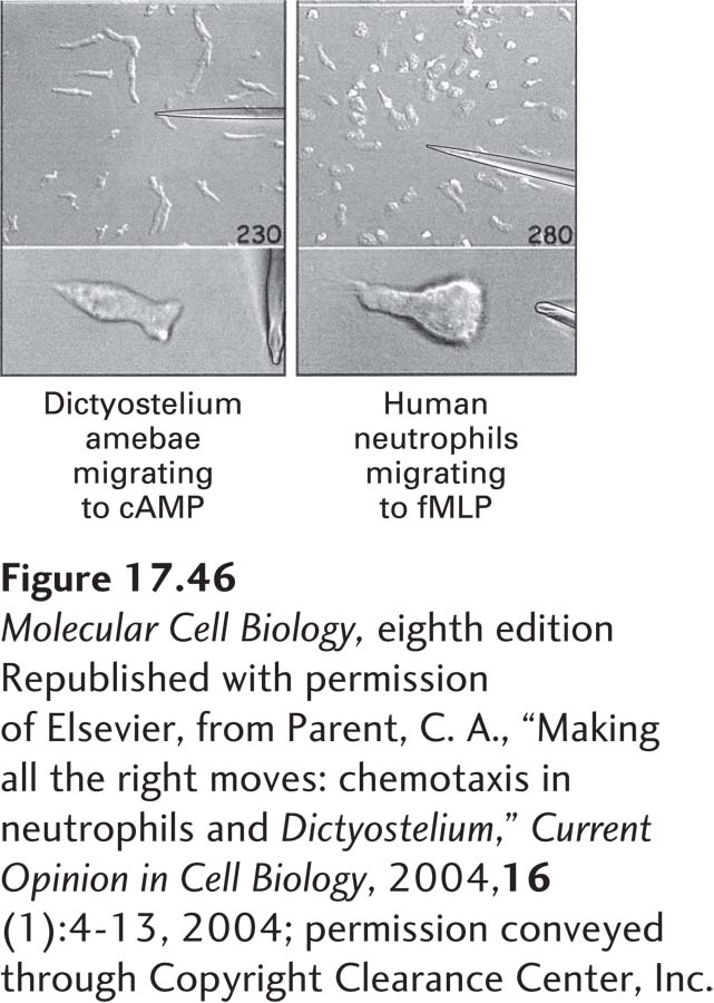
EXPERIMENTAL FIGURE 17-46 Chemotactic molecules guide migrating cells by signaling the actin cytoskeleton. Dictyostelium cells migrate toward a pipette of cAMP (left), and human neutrophils (a type of leukocyte) migrate toward a pipette of fMLP (formylated Met-Leu-Phe), a chemotactic peptide produced by bacteria (right). In the lower two panels are individual chemotaxing Dictyostelium and neutrophil cells, which look remarkably similar, despite about 800 million years of evolution separating them.
[Republished with permission of Elsevier, from Parent, C. A., “Making all the right moves: chemotaxis in neutrophils and Dictyostelium,” Current Opinion in Cell Biology, 2004, 16(1):4-13, 2004; permission conveyed through Copyright Clearance Center, Inc.]
Despite the variety of different chemotactic molecules—sugars, peptides, cell metabolites, cell-wall or membrane lipids—they all work through a common and familiar mechanism: binding to cell-surface receptors, activation of intracellular signaling pathways, and remodeling of the cytoskeleton through the activation or inhibition of various actin-binding proteins. What is quite amazing is that just a 2 percent difference in the concentration of chemotactic molecules between the front and back of the cell is sufficient to induce directed cell migration (see Classic Experiment 17-2). Equally amazing is the finding that the internal signal transduction pathways used in chemotaxis have been conserved between Dictyostelium amoebae and human leukocytes despite almost a billion years of evolution.