In the early days of electron microscopy, cell biologists noted long tubules in the cytoplasm that they called microtubules. Morphologically similar microtubules were seen making up the fibers of the mitotic spindle, as components of axons, and as the structural elements in cilia and flagella (Figure 18-2a, b). Careful examination of single microtubules from various sources in transverse section indicated that they are all made up of 13 longitudinal repeating units (Figure 18-2c), now called protofilaments, suggesting that these various microtubules all had a common structure. Microtubules purified from brain were then found to consist of a major protein, tubulin, and associated proteins, microtubule-associated proteins (MAPs). Purified tubulin alone can assemble into a microtubule under favorable conditions, proving that tubulin is the structural component of the microtubule wall. MAPs, as we will see, help mediate the assembly, dynamics, and function of microtubules. In this section, we consider the general structure and organization of microtubules before turning to a more detailed discussion of their dynamics and regulation in Sections 18.2 and 18.3.
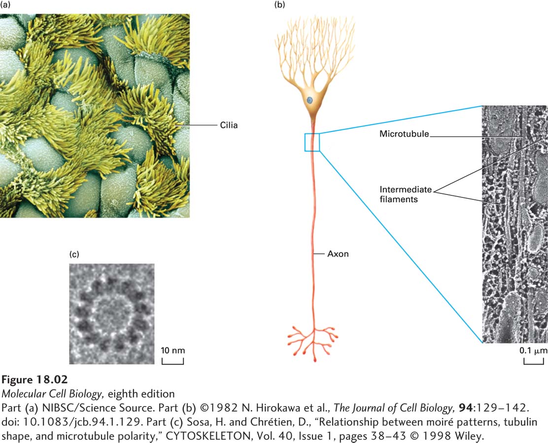
FIGURE 18-2 Microtubules are found in many different locations, and all have similar structures. (a) Surface of the ciliated epithelium lining a rabbit oviduct viewed in a scanning electron microscope. Beating cilia, each of which has a core of microtubules, propel eggs down the oviduct. (b) Microtubules and intermediate filaments in a quick-frozen and deep-etched frog axon visualized in a transmission electron microscope. (c) High-magnification view of a single microtubule showing the 13 repeating units known as protofilaments.
[Part (a) NIBSC/Science Source. Part (b) ©1982 N. Hirokawa et al., The Journal of Cell Biology, 94:129–142. doi: 10.1083/jcb.94.1.129. Part (c) Sosa, H. and Chrétien, D., “Relationship between moiré patterns, tubulin shape, and microtubule polarity,” CYTOSKELETON, Vol. 40, Issue 1, pages 38-43 © 1998 Wiley.]