Mitosis Can Be Divided into Six Stages
Mitosis has been divided up into several stages for ease of description (Figure 18-37a), but in reality it is a continuous process. Here we review the major events of each stage.
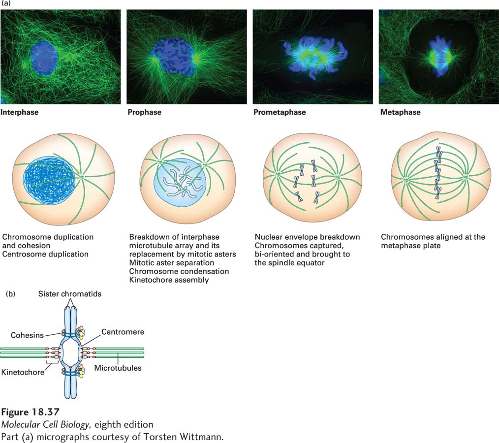
FIGURE 18-37 The stages of mitosis. (a) Upper panels show stages in cultured cells stained blue for DNA and green for tubulin. Lower diagrams show the different stages and the events that occur in each. Mitosis is a continuous process that has been divided into stages for ease of description. (b) Parts of a condensed chromosome in mitosis. The duplicated chromosome consists of two sister chromatids (each is a single DNA duplex), held together by cohesins at a constricted region called the centromere. The centromere is also the site where the kinetochore, which will make attachments to the kinetochore microtubules, forms.
[Part (a) micrographs courtesy of Torsten Wittmann.]
The first stage of mitosis, called prophase, is signaled by a number of coordinated and dramatic events. First, the interphase array of microtubules is replaced as the duplicated centrosomes become more active in microtubule nucleation. This activity provides two sites of assembly for dynamic microtubules, forming the mitotic asters. Additionally, the dynamics of the growing microtubules themselves increase due to changes in the activities of +TIPs at their (+) ends. The two asters are then moved to opposite sides of the nucleus by the action of bipolar kinesin-5 motors (see Figure 18-20) that push the intermingling microtubules apart. The separated centrosomes will become the two poles of the mitotic spindle, the microtubule-based structure that separates chromosomes. Second, protein synthesis is switched from being dependent to independent on the mRNA 5′ cap (see Section 5.4), and the internal membrane systems, normally dependent on the interphase array of microtubules, are disassembled. In addition, endocytosis and exocytosis are halted, and the microfilaments are generally rearranged to give rise to a rounded cell. In the nucleus, the nucleolus breaks down and chromosomes begin to condense. The cohesins holding together each pair of duplicated chromosomes, or sister chromatids as they are called at this stage, are degraded except at the centromeric region, where the two sister chromatids remain linked by intact cohesins (Figure 18-37b). Also during prophase, specialized structures called kinetochores, which will become sites of microtubule attachment, assemble at the centromeric region of each sister chromatid. As discussed in more detail in Chapter 19, all these events are coordinated by a rapid increase in the activity of the mitotic cyclin-CDK complex, which is a kinase that drives cell cycle progression by phosphorylating multiple proteins.
The next stage of mitosis, prometaphase, is initiated by the breakdown of the nuclear envelope and its retraction into the endoplasmic reticulum, the disassembly of nuclear pores, and the disassembly of the lamin-based nuclear lamina. Microtubules assembled from the spindle poles search for and “capture” chromosome pairs by associating with their kinetochores. Each chromatid has a kinetochore, so a sister chromatid pair has two kinetochores, each of which becomes attached through the microtubules to a different spindle pole during prometaphase in a critical process we discuss in detail below. The chromosome pairs then become aligned at a point equidistant between the two spindle poles. Prometaphase continues until all chromosomes have become aligned, at which point the cell enters the next stage, metaphase.
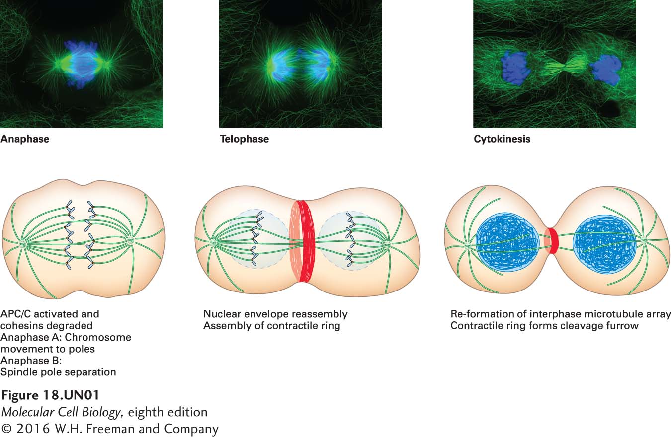
When the cell detects that all chromosomes are attached to the spindle correctly, the next stage, anaphase, is induced by activation of the anaphase-promoting complex or cyclosome (APC/C) (discussed in Chapter 19). The activated APC/C (through several intermediary steps) ultimately leads to the destruction of the cohesins that were holding the sister chromatids together, so that each chromatid can be pulled to its respective pole by the microtubules attached to its kinetochore. This movement is known as anaphase A. A separate and distinct movement also occurs: the movement of the spindle poles farther apart in a process known as anaphase B. Now that the chromosomes have separated, the cell enters telophase, when the nuclear envelope re-forms, the chromosomes decondense, and the cell is pinched into two daughter cells by the contractile ring during cytokinesis.