Anaphase A Moves Chromosomes to Poles by Microtubule Shortening
The onset of anaphase A is one of the most dramatic movements that can be observed in the light microscope. When the spindle assembly checkpoint has been passed, APC/C activation induces proteolysis of the remaining cohesins holding the sister chromatids together. Suddenly, the two paired sister chromatids separate from each other and are drawn to their respective poles. The movement is sudden because the kinetochore microtubules are under tension, and as soon as the cohesin attachments between the chromatids are removed, the separated chromatids are free to move.
Experiments with isolated metaphase chromosomes have shown that anaphase A movement can be powered by microtubule shortening, using the stored structural strain released by removal of the GTP-bound tubulin subunits at the microtubule tip. This mechanism can be nicely demonstrated in vitro. When metaphase chromosomes are added to purified microtubules, they bind preferentially to the (+) ends of the microtubules. Dilution of the mixture to reduce the concentration of free tubulin dimers results in the movement of the chromosomes toward the (−) ends by microtubule depolymerization at the chromosome-bound (+) ends. In addition, recent experiments have shown that in Drosophila, two members of the microtubule-depolymerizing kinesin-13 protein family (see Figure 18-15) also contribute to chromosome movement in anaphase A. One of these kinesin-13 proteins is localized at the kinetochore and enhances disassembly there (Figure 18-43, A1), and the other is localized at the spindle pole, enhancing depolymerization there (Figure 18-43, A2). Thus, at least in the fly, anaphase A is powered in part by kinesin-13 proteins specifically localized at the kinetochore and spindle pole to shorten the kinetochore microtubules at both their (+) and (−) ends, drawing the chromosomes to the poles.
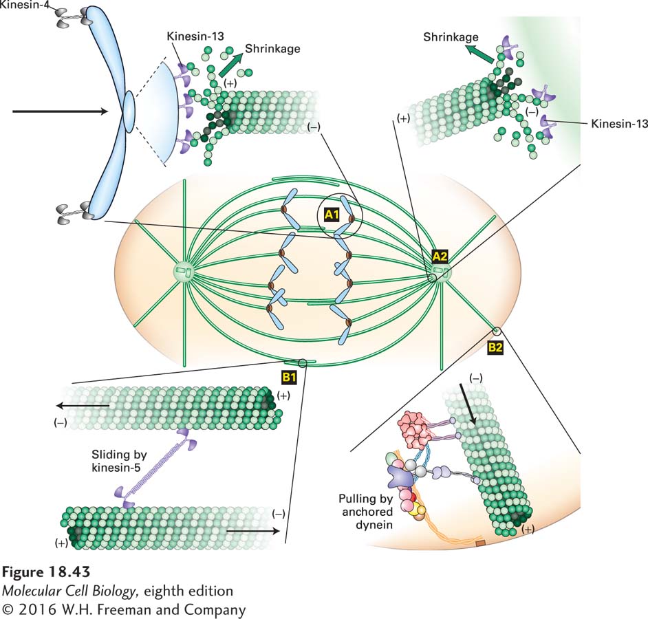
FIGURE 18-43 Chromosome movement and spindle pole separation in anaphase. Anaphase A movement is powered by microtubule-shortening kinesin-13 proteins at the kinetochore (A1) and at the spindle pole (A2). Note that the chromosome arms still point away from the spindle poles due to associated chromokinesin/kinesin-4 members, so the depolymerization force has to be able to overcome the force pulling the arms toward the center of the spindle. Anaphase B also has two components: sliding of antiparallel polar microtubules powered by a kinesin-5 (+) end–directed motor (B1), and pulling on astral microtubules by dynein-dynactin located at the cell cortex (B2). Arrows indicate the direction of movement generated by the respective forces. See Cleveland et al., 2003, Cell 112:407–421.