Cytokinesis Splits the Duplicated Cell in Two
During late anaphase and telophase in animal cells, the cell assembles a microfilament-based contractile ring attached to the plasma membrane that will eventually contract and pinch the cell into two, a process known as cytokinesis (see Figure 18-37). The contractile ring is a thin band of actin filaments of mixed polarity interspersed with myosin-II bipolar filaments (see Figure 17-35). On receiving a signal, the ring contracts, first to generate a cleavage furrow and then to pinch the cell into two.
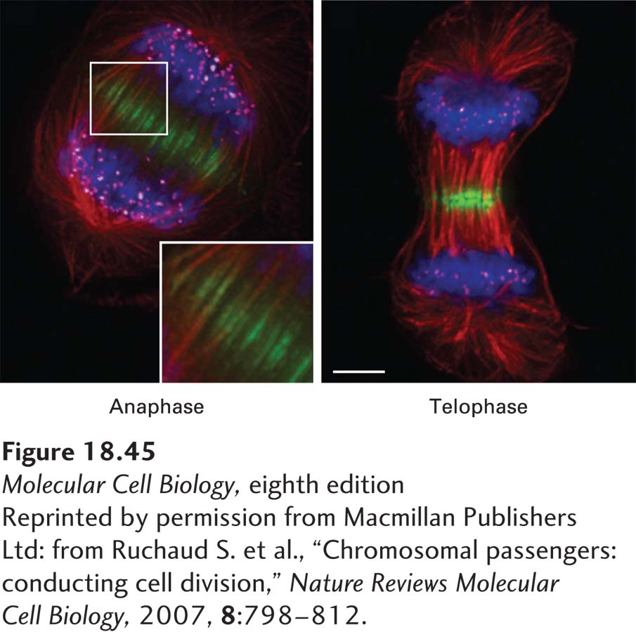
Two aspects of the contractile ring are essential to its function. First, it has to be appropriately placed in the cell. It is known that this placement is determined by a signal provided by the spindle, so that the ring forms equidistant between the two spindle poles. The signal is provided, at least in part, by the chromosomal passenger complex (CPC) that regulates the attachment of microtubules to kinetochores during prometaphase (see Figure 18-42b). Up to anaphase, the CPC is associated with the inner kinetochores of unseparated chromatids. When anaphase begins, it leaves the centromeres and associates with the overlapping polar microtubules at the center of the spindle (Figure 18-45). There the CPC recruits another protein complex, centralspindlin, that includes a (+) end–directed kinesin motor protein, which concentrates at the middle of the spindle due to its motor activity. As anaphase B continues, centralspindlin recruits a guanine nucleotide exchange factor for RhoA. Recall from Chapter 17 that Rho proteins are small GTP-binding proteins that are activated by exchange factors to catalyze the exchange of GDP for GTP (see Figure 17-41). Once activated, the RhoA⋅GTP activates a formin protein to drive the nucleation and assembly of actin filaments that make up the contractile ring (see Figure 17-43). In this way, the position of the spindle directly defines the site of contractile ring formation, and hence cytokinesis.

EXPERIMENTAL FIGURE 18-45 The chromosomal passenger complex (CPC) remains at the spindle midzone during anaphase and telophase. Micrographs of a cell in late anaphase (left) and telophase (right), showing microtubules (red), DNA (blue), Aurora B kinase (green), and kinetochores (magenta). Notice how the Aurora B, which is part of the CPC, concentrates in the region where the polar microtubules overlap and where the contractile ring will form. Scale bar 5 µm.
[Reprinted by permission from Macmillan Publishers Ltd: from Ruchaud S. et al., “Chromosomal passengers: conducting cell division,” Nature Reviews Molecular Cell Biology, 2007, 8:798–812.]
The second important aspect of the contractile ring is the timing of its contraction: if it were to contract before all chromosomes had moved to their respective poles, disastrous genetic consequences would ensue. As we discuss in Chapter 19, a signaling pathway has been discovered in budding yeast called the spindle position checkpoint, which pauses the cell cycle to ensure that cytokinesis does not occur until the spindle is appropriately oriented. The mechanism of this coordination in animal cells is still being unraveled.
