Interphase plant cells lack a central MTOC that organizes microtubules into the radiating array typical of animal cells. Instead, numerous MTOCs containing γ-tubulin line the cortex of plant cells and nucleate the assembly of transverse bands of microtubules below the cell wall (Figure 18-46, left). These microtubules, which are of mixed polarity, are released from the cortical MTOCs by the action of katanin, a microtubule-severing protein; loss of katanin gives rise to very long microtubules and misshapen cells. The reason for this is that these cortical microtubules, which are cross-linked by plant-specific MAPs, aid in laying down extracellular cellulose microfibrils, the main component of the rigid cell wall (see Figure 20-40).
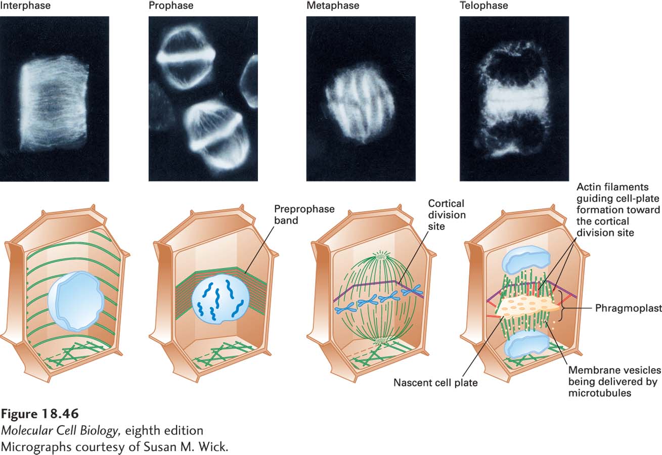
FIGURE 18-46 Mitosis in a flowering plant cell. Immunofluorescence micrographs (top) and corresponding diagrams (bottom) showing arrangement of microtubules in interphase and mitotic plant cells. A cortical array of microtubules girdles a cell during interphase. As the cell enters prophase, the microtubules (green), together with actin filaments (red), assemble under the cell cortex into a preprophase band, which marks the future cortical division site. As the cell enters prometaphase and metaphase, a spindle similar to that seen in animal cells forms. However, due to the cell wall, cytokinesis in plant cells is very different from that in in animal cells. Microtubules deliver vesicles whose membranes are used to assemble a membrane network called a phragmoplast, whose organization is defined by actin filaments linked to the cortical division site. Eventually, the phragmoplast becomes part of the plasma membranes of the two daughter cells. Enzymes secreted from the vesicles then build a cell wall between the two daughter cells. See G. Jürgens, 2005, Annu. Rev. Plant Biol. 56:281–299
[Micrographs courtesy of Susan M. Wick.]
Although mitotic events in plant cells are generally similar to those in animal cells, formation of the spindle and cytokinesis have unique features in plants (see Figure 18-46). Plant cells bundle their cortical microtubules and actin filaments into a preprophase band and reorganize them into a spindle at prophase without the aid of centrosomes. The site of the preprophase band defines the later cortical division site. At metaphase, the mitotic apparatus appears much the same in plant and animal cells. Because plants have cell walls, the division of the cell into two is quite different from animal cells and requires the assembly of a new wall between the daughter cells. Golgi-derived vesicles, which appear at telophase, are transported along microtubules to form the nascent cell plate. The cell plate expands and is guided toward the division site by actin filaments to form the phragmoplast, a membrane structure that replaces the animal-cell contractile ring. The membranes of the vesicles forming the phragmoplast become the plasma membranes of the daughter cells. The contents of these vesicles, such as polysaccharide precursors of cellulose and pectin, form the early cell plate, which develops into the new cell wall between the daughter cells.