The Nucleus Contains the DNA Genome, RNA Synthetic Apparatus, and a Fibrous Matrix
The nucleus, the largest organelle in animal cells, is surrounded by two membranes, each one a phospholipid bilayer containing many different types of proteins (Figure 1-15). The inner nuclear membrane defines the nucleus itself. In most cells, the outer nuclear membrane is continuous with the endoplasmic reticulum, and the space between the inner and outer nuclear membranes is continuous with the lumen of the endoplasmic reticulum (see Figure 1-15a). The two nuclear membranes appear to fuse at nuclear pore complexes, ringlike structures composed of specific membrane proteins through which material moves between the nucleus and the cytosol. The structure of the nuclear pores and the regulated transport of material through them are detailed in Chapters 10 and 13. Intermediate-filament proteins called lamins form a two-dimensional network, called the nuclear lamina, along the inner surface of the inner membrane, giving it shape and rigidity. The breakdown of the lamina occurs early in cell division, as we detail in Chapter 19. In a growing or differentiating cell, the nucleus is metabolically active, as it is the site of DNA replication and the synthesis of ribosomal RNA, mRNA, and a large variety of noncoding RNAs (see Chapters 5 and 9). Inside the nucleus one can often see a dense subcompartment, termed the nucleolus, where ribosomal RNA is synthesized and ribosomes are assembled (see Figure 1-15b and Chapter 10).
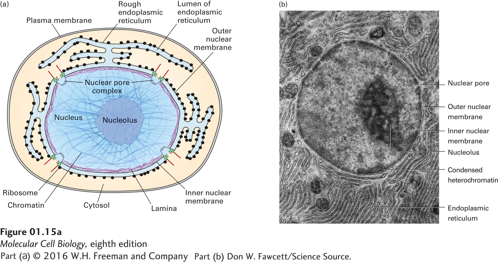
FIGURE 1-15 Structure of the nucleus. (a) Schematic diagram of the structure of a typical cell nucleus and the connection of the outer nuclear membrane with the rough endoplasmic reticulum. The small black dots attached to the membrane of the rough endoplasmic reticulum are ribosomes that are synthesizing membrane and secreted proteins. (b) Electron micrograph of a pancreatic acinar cell from the bat Myotis lucifugus. The nucleolus is a subcompartment of the nucleus and is not surrounded by a membrane; most ribosomal RNA is produced in the nucleolus. Darkly staining areas in the nucleus outside the nucleolus are regions of heterochromatin.
[Part (b) Don W. Fawcett/Science Source.]
The total DNA in an organism is referred to as its genome. In most prokaryotic cells, most or all of the genetic information resides in a single circular DNA molecule about a millimeter in length; this molecule lies, folded back on itself many times, in the central region of the micrometer-sized cell (see Figure 1-11). In contrast, DNA in the nuclei of eukaryotic cells is distributed among multiple long linear structures called chromosomes. The length and number of chromosomes are the same in all cells of a particular species, but vary among different species (see Table 1-2). Each chromosome comprises a single DNA molecule associated with numerous histones and other proteins. In a nucleus that is not dividing, the chromosomes are dispersed and are not dense enough to be observed in the light microscope. Only during cell division are individual chromosomes visible by light microscopy. When nondividing cells are visualized in an electron microscope, the non-nucleolar regions of the nucleus, called the nucleoplasm, can be seen to have dark- and light-staining areas. The dark areas, which are often closely associated with the nuclear membrane, contain condensed, concentrated DNA that cannot be transcribed into RNA, called heterochromatin (see Figure 1-15b).
Chromosomes, which stain intensely with basic dyes, are visible in light and electron microscopes only during cell division, when the DNA becomes tightly compacted (Figure 1-16). Although the large genomic DNA molecule in prokaryotes is associated with proteins, the arrangement of DNA within a bacterial chromosome differs greatly from that within the linear chromosomes of eukaryotic cells; bacterial chromosomes are circular and are associated with different types of proteins than are eukaryotic chromosomes.
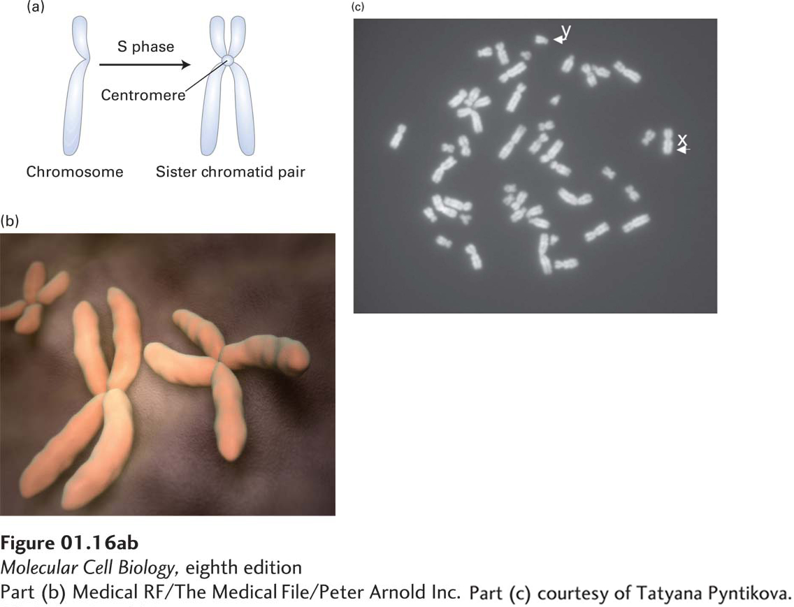
FIGURE 1-16 Individual chromosomes can be seen in cells during cell division (mitosis). (a) During the S phase of the cell cycle (see Figure 1-21) chromosomes are duplicated, and the daughter “sister chromatids,” each with a complete copy of the chromosomal DNA, remain attached at the centromere. (b) During the actual cell division process (mitosis), the chromosomal DNA becomes highly compacted, and the pairs of sister chromatids can be seen in the electron microscope, as depicted here. (c) Light-microscope image of a chromosomal spread from a cultured human male lymphoid cell arrested in the metaphase stage of mitosis by treatment with the microtubule-depolymerizing drug colcemid. There is a single copy of the duplicated X and Y chromosomes and two copies of each of the others.
[Part (b) Medical RF/The Medical File/Peter Arnold Inc. Part (c) courtesy of Tatyana Pyntikova.]