Tissues Are Organized into Organs
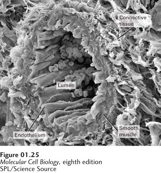
FIGURE 1-25 All organs are organized arrangements of various tissues, as illustrated in this cross section of a small artery (arteriole). Blood flows through the vessel lumen, which is lined by a thin sheet of endothelial cells forming the endothelium and by the underlying basal lamina. This tissue adheres to the overlying layer of smooth muscle tissue; contraction of the muscle layer controls blood flow through the vessel. A fibrillar layer of connective tissue surrounds the vessel and connects it to other tissues.
[SPL/Science Source.]
Cells in metazoans do not work in isolation; specialized groups of differentiated cells often form tissues, which are themselves the major components of organs. For example, the lumen of a small blood vessel is lined with a sheet-like layer of endothelial cells, or endothelium, which prevents blood cells from leaking out (Figure 1-25). A layer of smooth muscle tissue encircles the endothelium and basal lamina and contracts to limit blood flow. During times of fright, constriction of smaller peripheral vessels forces more blood to the vital organs. The muscle layer of a blood vessel is wrapped in an outer layer of connective tissue, a network of fibers and cells that encases the vessel walls and protects them from stretching and rupture.
This hierarchy of tissues is copied in other blood vessels, which differ mainly in the thickness of the layers. The wall of a major artery must withstand much stress and is therefore thicker than that of a minor vessel. The strategy of grouping and layering different tissues is used to build other complex organs as well. In each case, the function of the organ is determined by the specific functions of its component tissues, and each type of cell in a tissue produces the specific groups of proteins that enable the tissue to carry out its functions.