22.2 Voltage-Gated Ion Channels and the Propagation of Action Potentials
In Chapter 11 we learned that an electric potential of ~70 mV (cytosolic face negative) exists across the plasma membrane of all cells, including resting nerve cells. This resting membrane potential is generated by outward movement of K+ ions through open nongated K+ channels in the plasma membrane, and is driven by the K+ concentration gradient (cytosol > extracellular medium). The high cytosolic K+ and low cytosolic Na+ concentrations, relative to their concentrations in the extracellular medium, are generated by the plasma membrane Na+/K+ pump, which uses the energy released by hydrolysis of phosphoanhydride bonds in ATP to pump Na+ outward and K+ inward. The entry of Na+ ions into the cytosol from the medium is thermodynamically favored, driven both by the Na+ concentration gradient (extracellular medium > cytosol) and the inside-negative membrane potential (see Figure 11-25). However, most Na+ channels in the plasma membrane are closed in resting cells, so little inward movement of Na+ ions can occur (Figure 22-9a).
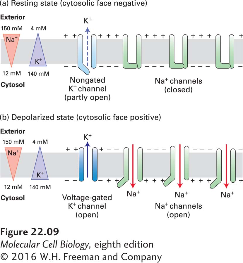
FIGURE 22-9 Depolarization of the plasma membrane due to opening of gated Na+ channels. (a) In resting neurons, a type of nongated K+ channel is open part of the time, but the more numerous gated Na+ channels are closed. The movement of K+ ions outward establishes the inside-negative membrane potential characteristic of most cells. (b) Opening of gated Na+ channels permits an influx of sufficient Na+ ions to cause a reversal of the membrane potential. In the depolarized state, voltage-gated K+ channels open and subsequently repolarize the membrane. Note that the flows of ions are too small to have much effect on the overall concentration of either Na+ or K+ in the cytosol or exterior fluid.
During an action potential, some of these Na+ channels open, allowing inward movement of Na+ ions, which depolarizes the membrane. Action potentials are propagated down the axon because a change in voltage in one part of the axon triggers the opening of channels in the next section of the axon. Such voltage-gated channels therefore lie at the heart of neural transmission. In this section, we first introduce some of the key properties of action potentials, which move rapidly along the axon from the cell body to the termini. We then describe how the voltage-gated channels responsible for propagating action potentials in neurons operate. In the last part of the section, we will see how the myelin sheath, produced by glial cells, increases the speed and efficiency of electrical transmission in nerve cells.