Viral Capsids Are Regular Arrays of One or a Few Types of Protein
The nucleic acid of a virion is enclosed within a protein coat, or capsid, composed of multiple copies of one protein or a few different proteins, each of which is encoded by a single viral gene. Because of this structure, a virus is able to encode all the information for making a relatively large capsid in a small number of genes. This efficient use of genetic information is important because only a limited amount of DNA or RNA, and therefore a limited number of genes, can fit into a virion capsid.
Nature has found two basic ways of arranging the multiple capsid protein subunits and the viral genome into a virion. In some viruses, multiple copies of a single capsid protein form a helical structure that encloses and protects the viral RNA or DNA, which runs in a helical groove within the protein tube. Viruses with such a helical structure, such as tobacco mosaic virus, have a rodlike shape (Figure 5-43a). The other major structural type is based on the icosahedron, a solid, approximately spherical object built of 20 identical faces, each of which is an equilateral triangle (Figure 5-43b). During infection, some icosahedral viruses interact with host-
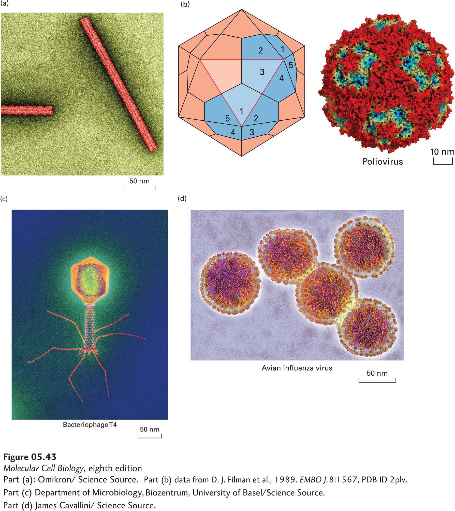
In many DNA bacteriophages, the viral DNA is located within an icosahedral “head” that is attached to a rodlike “tail.” During infection, viral proteins at the tip of the tail bind to host-
In some viruses, a symmetrically arranged nucleocapsid composed of the viral genome associated with multiple copies of one or a few proteins is covered by an external membrane, or envelope, which consists mainly of a phospholipid bilayer but also contains one or more types of virus-