Chapter Introduction
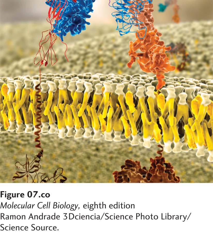
Molecular model of a lipid bilayer with embedded membrane proteins. Integral membrane proteins have distinct exoplasmic, cytosolic, and membrane-spanning domains. Shown here are portions of the insulin receptor, which regulates cell metabolism.
[Ramon Andrade 3Dciencia/Science Photo Library/Science Source.]
OUTLINE
7.1 The Lipid Bilayer: Composition and Structural Organization
7.2 Membrane Proteins: Structure and Basic Functions
7.3 Phospholipids, Sphingolipids, and Cholesterol: Synthesis and Intracellular Movement
Membranes participate in many aspects of cell structure and function. The plasma membrane defines the cell and separates the inside from the outside. In eukaryotes, membranes also define intracellular organelles such as the nucleus, mitochondrion, and lysosome. These biomembranes all have the same basic architecture: a phospholipid bilayer in which proteins are embedded (Figure 7-1). By preventing the unassisted movement of most water-soluble substances across the membrane, the phospholipid bilayer serves as a permeability barrier, helping to maintain the characteristic differences between the inside and the outside of the cell or organelle; in turn, the embedded proteins endow the membrane with specific functions, such as regulated transport of substances from one side to the other. Each cellular membrane has its own set of proteins that allow it to carry out many different functions.
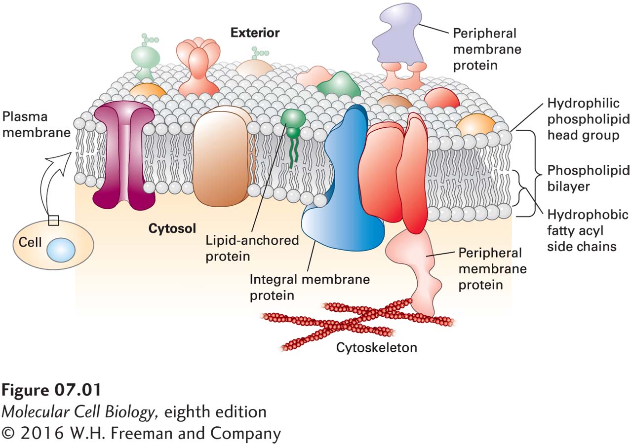
FIGURE 7-1 Fluid mosaic model of biomembranes. A bilayer of phospholipids about 3 nm thick provides the basic architecture of all cellular membranes; membrane proteins give each cellular membrane its unique set of functions. Individual phospholipids can move laterally and spin within the plane of the membrane, giving the membrane a fluidlike consistency similar to that of olive oil. Noncovalent interactions between phospholipids, and between phospholipids and proteins, lend strength and resilience to the membrane, while the hydrophobic core of the bilayer prevents the unassisted movement of water-soluble substances from one side to the other. Integral membrane proteins (transmembrane proteins) span the bilayer and often form dimers and higher-order oligomers. Lipid-anchored proteins are tethered to one leaflet by a covalently attached hydrocarbon chain. Peripheral proteins associate with the membrane primarily by specific noncovalent interactions with integral membrane proteins or membrane lipids. Proteins in the plasma membrane also make extensive contact with the cytoskeleton. See D. Engelman, 2005, Nature 438:578–580.
Prokaryotes, the simplest and smallest cells, are about 1–2 µm in length and are surrounded by a single plasma membrane; in most cases, they contain no internal membrane-limited subcompartments (see Figure 1-11). However, this single plasma membrane contains hundreds of different types of proteins that are integral to the function of the cell. Some of these proteins catalyze ATP synthesis and initiation of DNA replication, for instance. Others include the many types of membrane transport proteins that enable specific ions, sugars, amino acids, and vitamins to cross the otherwise impermeable phospholipid bilayer to enter the cell and that allow specific metabolic products to exit. Receptor proteins in the plasma membrane are proteins that allow the cell to recognize chemical signals present in its environment and adjust its metabolism or pattern of gene expression in response.
Eukaryotes also have a plasma membrane studded with many different proteins that perform a variety of functions, including membrane transport, cell signaling, and connecting cells into tissues. In addition, eukaryotic cells—which are generally much larger than prokaryotes—have a variety of internal membrane-bounded organelles (see Figure 1-12). Each type of organelle membrane has a unique complement of proteins that enable it to carry out its characteristic cellular functions, such as ATP generation (in mitochondria) and DNA synthesis (in the nucleus). Many plasma-membrane proteins also bind components of the cytoskeleton, a dense network of protein filaments that crisscrosses the cytosol to provide mechanical support for cellular membranes. These interactions are essential for the cell to assume its specific shape and for many types of cell movements.
Despite playing a structural role in cells, membranes are not rigid structures. They can bend and flex in three dimensions while still maintaining their integrity, in part because of the abundant noncovalent interactions that hold the lipids and proteins together. Moreover, there is considerable mobility of individual lipids and proteins within the plane of the membrane. According to the fluid mosaic model of biomembranes, first proposed by researchers in the 1970s, the phospholipid bilayer behaves in some respects like a two-dimensional fluid, with individual lipid molecules able to move past one another as well as spin in place. Such fluidity and flexibility not only allows organelles to assume their typical shapes, but also confers on the membrane the dynamic property that enables membrane budding and fusion, as occurs when viruses are released from an infected cell (Figure 7-2a) or when the internal cellular membranes of the Golgi complex bud into vesicles in the cytosol (Figure 7-2b) and then fuse with other membranes to transport their contents from one organelle to another (see Chapter 14).
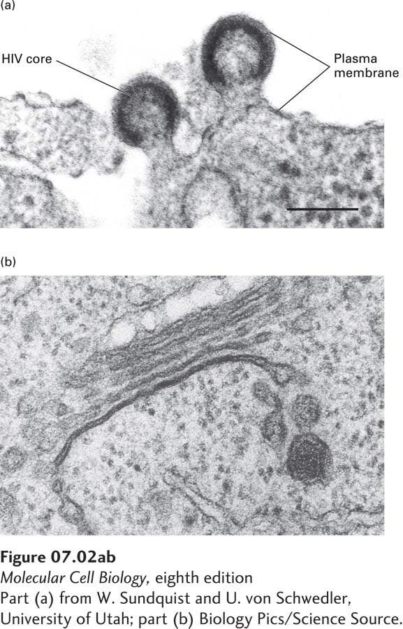
FIGURE 7-2 Eukaryotic cellular membranes are dynamic structures. (a) An electron micrograph of the plasma membrane of an HIV-infected cell, showing HIV particles budding into the culture medium. As the virus core buds from the cell, it becomes enveloped by a membrane, derived from the cell’s plasma membrane, that contains specific viral proteins. (b) Stacked membranes of the Golgi complex with budding vesicles. Note the irregular shape and curvature of these membranes.
[Part (a) from W. Sundquist and U. von Schwedler, University of Utah; part (b) Biology Pics/Science Source.]
We begin our examination of biomembranes by considering their lipid components. These molecules not only affect membrane shape and function, but also help anchor proteins to the membrane, modify membrane protein activities, and transduce signals to the cytoplasm. We then consider the structure of membrane proteins. Many of these proteins have large segments that are embedded in the hydrocarbon core of the phospholipid bilayer, and we focus on the principal classes of such transmembrane proteins. Finally, we consider how lipids such as phospholipids and cholesterol are synthesized in cells and distributed to their many membranes and organelles.