Fatty Acids Are Incorporated into Phospholipids Primarily on the ER Membrane
Fatty acids are not directly incorporated into phospholipids; rather, in eukaryotic cells, they are first converted into CoA esters. The subsequent synthesis of phospholipids such as the phosphoglycerides is carried out by enzymes associated with the cytosolic face of the ER membrane, usually the smooth ER, in animal cells; through a series of steps, fatty acyl CoAs, glycerol 3-phosphate, and polar head group precursors are linked together and then inserted into the ER membrane (Figure 7-25). The fact that the enzymes involved in this process are located on the cytosolic side of the membrane means that there is an inherent asymmetry in membrane biogenesis: new membranes are initially synthesized only on one leaflet—a fact with important consequences for the asymmetric distribution of lipids in membrane leaflets. Once synthesized on the ER, phospholipids are transported to other organelles and to the plasma membrane. Mitochondria synthesize some of their own membrane lipids and import others.
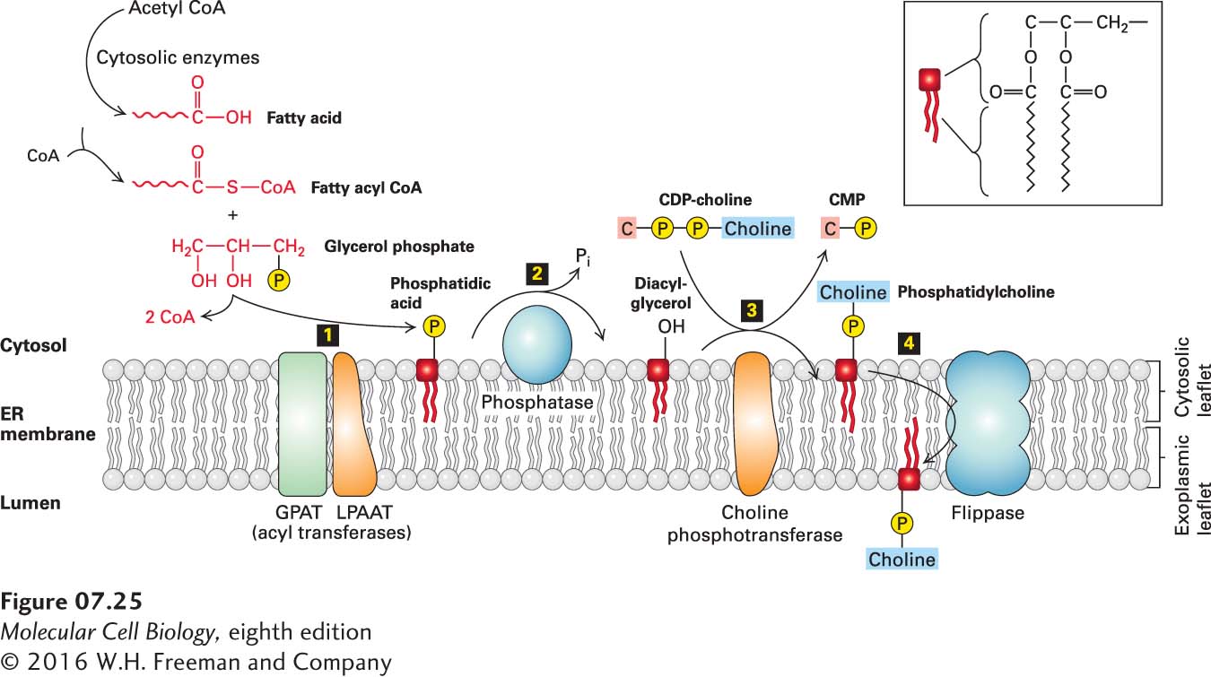
FIGURE 7-25 Phospholipid synthesis in the ER membrane. Because phospholipids are amphipathic molecules, the last steps of their multistep synthesis take place at the interface between a membrane and the cytosol and are catalyzed by membrane-associated enzymes. Step 1: Two fatty acids from fatty acyl CoA are esterified to the phosphorylated glycerol backbone, forming phosphatidic acid, whose two long hydrocarbon chains anchor the molecule to the membrane. Step 2: A phosphatase converts phosphatidic acid into diacylglycerol. Step 3: A polar head group (e.g., phosphorylcholine) is transferred from cytosine diphosphocholine (CDP-choline) to the exposed hydroxyl group. Step 4: Flippase proteins catalyze the movement of phospholipids from the cytosolic leaflet in which they are initially formed to the exoplasmic leaflet.
Sphingolipids are also synthesized indirectly from multiple precursors. Sphingosine, the building block of these lipids, is made in the ER, beginning with the coupling of a palmitoyl group from palmitoyl CoA to serine; the subsequent addition of a second fatty acyl group to form N-acyl sphingosine (ceramide) also takes place in the ER. Later, in the Golgi, a polar head group is added to ceramide, yielding sphingomyelin, whose head group is phosphorylcholine, and various glycosphingolipids, in which the head group may be a monosaccharide or a more complex oligosaccharide (see Figure 7-8b). Some sphingolipid synthesis can also take place in mitochondria. In addition to serving as the backbone for sphingolipids, ceramide and its metabolic products are important signaling molecules that can influence cell growth, proliferation, endocytosis, resistance to stress, and programmed cell death (apoptosis).
After their synthesis is completed in the Golgi, sphingolipids are transported to other cellular compartments through vesicle-mediated mechanisms similar to those for the transport of proteins, discussed in Chapter 14. Any type of vesicular transport results in movement not only of the protein payload, but also of the lipids that compose the vesicular membrane. Phospholipids such as phosphoglycerides, as well as cholesterol, can move between organelles by additional mechanisms, described below.