During Metaphase, Chromosomes Can Be Distinguished by Banding Patterns and Chromosome Painting
Certain dyes selectively stain some regions of metaphase chromosomes more intensely than other regions, producing characteristic banding patterns that are specific for individual chromosomes. The regularity of chromosome bands provides useful visible landmarks along the length of each chromosome and can help to distinguish chromosomes of similar size and shape, as we will see later in this section.
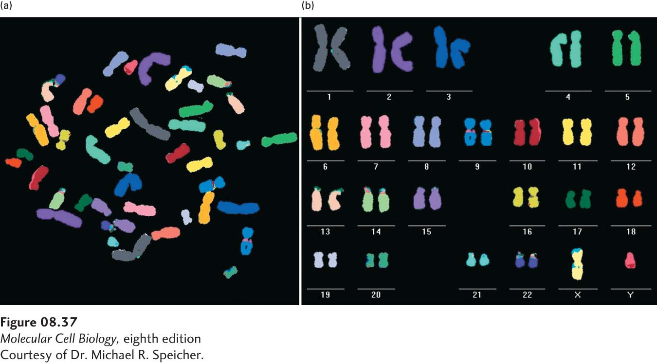
Today the method of chromosome painting greatly simplifies the identification and differentiation of individual chromosomes within a karyotype, many of which have similar sizes and shapes. This technique, a variation of fluorescence in situ hybridization (FISH), makes use of probes specific for sites scattered along the length of each chromosome. The probes are labeled with several different fluorescent dyes with distinct excitation and emission wavelengths. Probes specific for each chromosome are labeled with a predetermined fraction of each of the dyes. After the probes are hybridized to chromosomes and the excess removed, the sample is observed with a fluorescence microscope in which a detector determines the fraction of each dye present at each fluorescing position in the microscopic field. This information is conveyed to a computer, and a special program assigns a false-
342
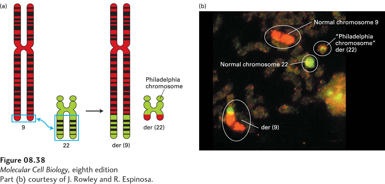
Chromosome painting is a powerful method for detecting an abnormal number of chromosomes, such as chromosome 21 trisomy in patients with Down syndrome, or chromosomal translocations that occur in rare individuals and in cancer cells (Figure 8-38). The use of probes with different ratios of fluorescent dyes that hybridize to distinct positions along each normal human chromosome allows finer structural analysis of the chromosomes that can more readily reveal deletions or duplications of chromosomal regions. The chapter-