Chapter 18. AKT1 and Vimentin
Introduction

Analyze the Data 18-1: AKT1 and Vimentin
The PI3K/AKT signaling pathway is aberrant in a wide variety of cancers. In soft-tissue sarcoma (STS) cells, activation of the kinase AKT1 induces cell motility and invasiveness, which leads to aggressive metastasis of the cells. AKT1 has been shown to bind to vimentin (see Q.-S. Zhu et al., 2011, Oncogene 30:457–470).
Question
a. To map the vimentin and AKT interaction domains, the vimentin and AKT1 full-length and fragment constructs indicated below were expressed as GST-fusion proteins. Each of the GST-fusion constructs bound to glutathione beads was used to recover associated proteins from crude STS-cell lysates. What did the Western blot analysis of the material recovered by GST fused to domains of vimentin using an AKT1 antibody reveal about the AKT-binding domain in vimentin? What did analysis of the material recovered by GST fused to AKT1 domain using a vimentin antibody reveal about the vimentin- binding domain in AKT1?
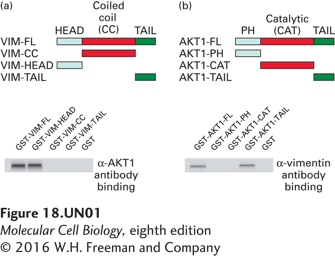
Both the full-length (GST-VIM-FL) vimentin and the vimentin head domain (GST-VIM-Head) GST fusion proteins pull down AKT1, but the vimentin coiled-coil domain (GST-VIM-CC) and the tail (GST-VIM-TAIL) regions do not (left). The full length AKT1 and the AKT1 tail domain pull down vimentin, but the PAH domain (GST-AKT1-PH) and the catalytic (GST-AKT1-CAT) domains do not. This demonstrates that the vimentin head domain interacts directly with the AKT1 tail domain.
Question
b. AKT1 is a kinase and is therefore likely to phosphorylate vimentin. Sequence analysis revealed that serines (S) at positions 39 and 325 in vimentin were likely sites for AKT1 phosphorylation. To test whether one or both of these sites is phosphorylated by AKT1, each site was mutated to alanine (A), which cannot be phosphorylated. Each alanine-mutated vimentin was mixed with AKT1 and tested for phosphorylation by Western blot analysis using an antibody that reacts with AKT1- phosphorylated serines (PAS antibody). Which site(s) does AKT1 phosphorylate?
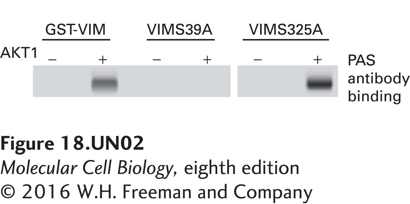
Western blot analysis with the PAS antibody that reacts with sites phosphorylated by AKT1 demonstrates that AKT1 phosphorylates the wild type and S325A mutated vimentins but not the S39A vimentin mutation. This identifies vimentin Serine 39 as the site of AKT1 phosphorylation.
Question
c. The propensity of cancer cells to metastasize can be measured with an invasion assay using a filter coated with extracellular matrix (ECM) proteins. The more cells that migrate through the filter, the higher the propensity for metastasis. The invasion assay was used to monitor the effects of expression of a variant of AKT1 with a mutation that makes the kinase constitutively active (AKT1DD); of overexpression of wild-type vimentin; of expression of a vimentin harboring a mutation so that it cannot be phosphorylated by AKT1 (VIMS39A); and of expression of a phosphomimetic vimentin mutation (VIMS39D) in which mutation of S39 to an aspartate residue (D) mimics phosphorylation of the serine. What effect does vimentin overexpression and phosphorylation have on cell migration?
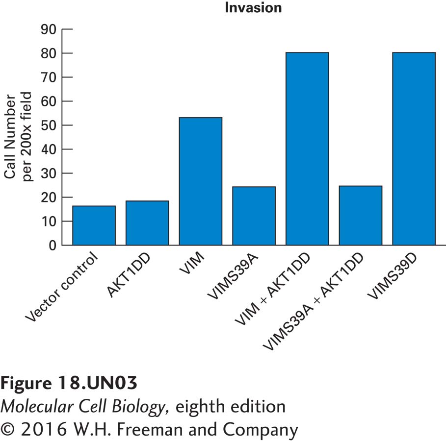
Expression of active AKT1 (AKT1DD) has no affect on cell migration, but over-expression of vimentin alone (VIM) significantly increases cell migration and that migration is further enhanced by expressing both vimentin and active AKT1 (VIM+AKT1DD). This enhancement is blocked by substituting the wild-type vimentin with the mutation that cannot be phosphorylated by AKT1 (VIMS39A +AKT1DD). Expression of the phosphomimetic form of the vimentin (VIMS39D), even in the absence of AKT1, mimics the effect of AKT1 phosphorylation of wild-type vimentin. These results indicate that phosphorylation of vimentin S39 or mimicking that phosphorylation maximizes cell migration.
Activity results are being submitted...