Chapter 23. Monitoring Antigen Presentation and T-Cell Activation
Introduction

Analyze the Data 23-1: Monitoring Antigen Presentation and T-Cell Activation
To understand how ovalbumin (a protein from chicken eggs) and other foreign antigens in the cytoplasm of a cell are presented for immunosurveillance, ovalbumin can be introduced by electroporation into the cytoplasm of mouse B lymphocytes in primary cell culture. The ovalbumin is cleaved, and one of its cleavage products is a peptide with the sequence SIINFEKL (single-letter abbreviations). When the B cells are mixed with a clonal T-cell population that specifically recognizes SIINFEKL in the context of MHC molecules, the T cells become stimulated. The graphs below show the secretion of IL-2 after these T cells were mixed with B cells that had been treated in various ways.
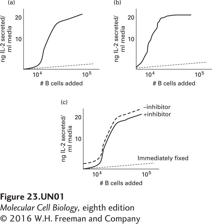
Graph A: B cells were electroporated either with ovalbumin (solid line) or with a control protein (dashed line) not recognized by the clonal T-cell population.
Graph B: B cells were first incubated with an inhibitor of lysosomal cysteine proteases (solid line) or an inhibitor of the proteasome (dashed line), then electroporated with ovalbumin.
Graph C: B cells were exposed to a high concentration of the SIINFEKL peptide and then immediately fixed [killed] (dotted line) with formaldehyde or incubated for 2 hours in the presence (dashed curve) or absence (solid curve) of the proteasome inhibitor and then fixed with formaldehyde.
Question
a1qkxiBtG+AoeuVJ+Qlo8R+7mvTNu2MXwWwgVA7V4X1o12TZui0qf6P+JVtEZrmHkCcPh8MphPoNZ4AlOELUi512VH/I/K9vN39VsHGTkIYquEBTXQyEePq/reEQtqFiGRyhnR2MINm+O/54l2AKoARDTlZt9w5b2xqmtMfNTooPB4UiZmLj1t1ees7OTvTGFdE7PA==Because, as shown in graph C, IL-2 is secreted in panel C and the B cells are killed, secretion of IL-2 must be from the T cells and would occur when the T cells are activated by the B cells. Activation would occur when the T cells recognize the SIINFEKL peptide, as the cells electroporated with the control protein (graph A) do not induce T-cell activation. Interestingly, the T cells are activated when SIINFEKL is presented to them on the surface of fixed cells (graph C, dashed and solid curves). Thus, B cells need not be alive, they just need to properly present antigen.
Question
5e7U35SsqHx4ZOKYNDYi0c6WpwCfq8ZcKznY3/yChXO2jcqCB2hgxmtZfKGAKIG/BIPRuiTX7nXbSJNyq9wFSqJBOWzeZfnheDMrDVfBobyH/S/W4G4KZGjO+4GOs9qlb+dJA1x71W9Z/WBVSKa8oEZ/YLzvb0tqciAhJ4xHNEldT6mXARc+FzUa+H9IOQmQtjWEQ+WbZiwDv6TSJmRXZwW835vqvW8I0zKH2sAcUea4bhA7sQToBfJn02FQZ9+DNfGmy0Rd2HZBQ96duOJUuWQHZF68pf8AwUTvwgnvfJkz3aFBksF+Z2T0W1yMfVHu1tcFRSGmzlMJst3/NLZKsiHi3CGeHsbuT6aCKLPOsBhMbH99c+e6eq6GdzvWrZHGHeoUBhZNJqAbXuNweszfLSonYXacCwP5zvp5qkhrUZsADYXdL9hEysrKLmEGuon6LbqsxpeEx8DdVIgFOELs2DgwPFxIOzHmBAVHJqg6oEWcOjtROne of the inhibitors blocks proteolysis in lysosomes, the other blocks proteolysis induced by proteasomes in the cytoplasm. Presentation by Class I MHC involves degradation of self and foreign molecules in the cytosol by proteasomes, whereas presentation by Class II MHC involves endocytosis of microbial pathogens, which are then degraded in phagosomes/lysosomes for anti-gen presentation. Ovalbumin, introduced into the cytoplasm of the cells, would be expected to be cleaved by the proteasome and its peptides trans-located into the ER for presentation by Class I MHC molecules. The Class I MHC-peptide complex would be transported via the secretory pathway to the cell surface. Thus, the absence of a T-cell response (graph B) in the presence of the proteasome inhibitor, but not the lysosomal inhibitor, suggests that ovalbumin follows the Class I MHC pathway, and not the Class II MHC pathway.
Question
r4CUWdt4avhOa46As7ITL10RlVZbYtom/HEOo8J8gXX/7PjfUzq4ETM/ofFVQRsDY+9v5HHkmBO9TWaXsNxrEKUMLISlj9e7fqHKedNNFrQN5ymo/jqT1ck7F6ry/7wlJ9HIzu6SdHTYFio+OKYX4eXO51PopeZXsJKV3M2zxqG2NXtfYcNt6Fmnr8tMk7y30VLBCsW6INXQ+Xv+y623ghDZImwBpcGjyiIbvpAUklCobC2lk0iVl/sHJqyWoJsAkMVrICmIGR3T1TpjF1sp/ZcgmhoqCyuDuQyaYYA8cw/23Rlriq/IIbD0We9eJby+7hxGJVcwLGxencH1fUvFDA==In the experiments shown in graph C, the SIINFEKL peptide, rather than intact ovalbumin, has been introduced into the B cells. Accordingly, there would be no need for the proteasome to digest ovalbumin; the appropriate peptide is already present. These data indicate that inhibition of the proteasome does not cause a deficiency in the cell that prevents peptides from being presented at the cell surface. In this case, SIINFEKL is presented regardless of proteasome function. Accordingly, the proteasome must digest the protein for antigen presentation.
Activity results are being submitted...