HOW DO WE KNOW?
FIG. 35.11
What is the resting membrane potential? How does electrical activity change during an action potential?
BACKGROUND In order to record the voltage of the inside of a nerve cell relative to the outside, a small electrical recording device called a microelectrode can be inserted into a neuron. The technique is more easily performed on large cells, such as the squid giant axon. This axon, as its name suggests, is quite large, measuring 0.5 mm in diameter (Fig. 35.11a). The large diameter of the axon allows electrical signals to be propagated to the muscles very quickly. Vertebrate neurons are much smaller; they rely on myelin sheaths rather than large size for rapid conduction of electrical signals.
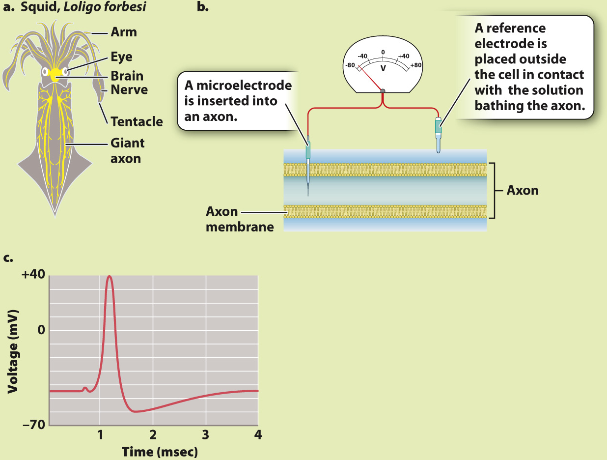
EXPERIMENT In 1939, two British neurophysiologists, Alan Hodgkin and Andrew Huxley, inserted a microelectrode into a squid giant axon and placed a reference electrode on the outside (Fig. 35.11b). They then used a separate set of electrodes (not shown) to depolarize the cell to threshold, triggering an action potential.
RESULTS Fig. 35.11c is a trace from Hodgkin and Huxley’s 1939 paper showing the resting membrane potential and the course of an action potential recorded by the electrode inside a giant squid axon. Note that the resting potential (–45 mV in squid) is negative on the inside of the axon relative to the outside, and that the action potential is a rapid spike in potential, with the inside of the cell quickly becoming positive, then negative again. The large size of the spike was a surprise.
FOLLOW-
SOURCES Hodgkin, A. L., and A. F. Huxley. 1939. “Action Potentials Recorded from Inside a Nerve Fibre.” Nature 144:710–