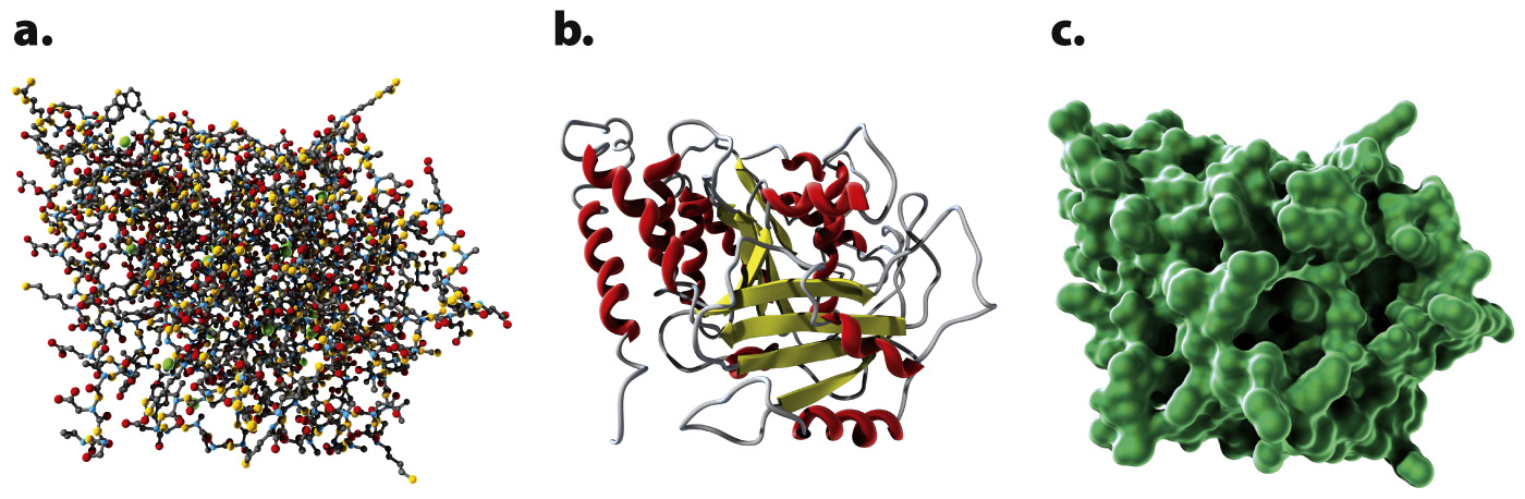
FIG. 4.8
Three ways of showing the structure of the protein tubulin:
(a) bal
l-
an
d-
stick model; (b) ribbon model; (c) spac
e-
filling model.