Microtubules and microfilaments are polymers of protein subunits.
Microtubules are hollow tubelike structures with the largest diameter of the three cytoskeletal elements, about 25 nm (Fig. 10.3a). They are polymers of protein dimers. Each dimer is made up of two slightly different tubulin proteins, called α (alpha) and β (beta) tubulin. One α tubulin and one β tubulin combine to make a tubulin dimer and the tubulin dimers are assembled to form the microtubule.
Microtubules help maintain cell shape and the cell’s internal structure. In animal cells, microtubules radiate outward to the cell periphery from a microtubule organizing center called the centrosome. This spokelike arrangement of microtubules helps cells withstand compression and thereby maintain their shape. Many organelles are tethered to microtubules, and thus microtubules guide the arrangement of organelles in the cell.
Microfilaments are polymers of actin monomers, arranged to form a helix. They are the thinnest of the three cytoskeletal fibers, about 7 nm in diameter, and are present in various locations in the cytoplasm (Fig. 10.3b). They are relatively short and extensively branched in the cell cortex, the area of the cytoplasm just beneath the plasma membrane. At the cortex, microfilaments reinforce the plasma membrane and organize proteins associated with it.
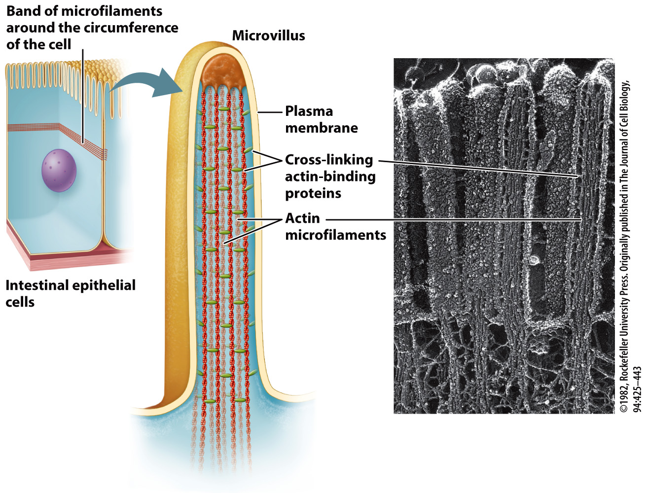
These cortical microfilaments are also important in part for maintaining the shape of a cell, such as the biconcave shape of red blood cells discussed earlier (see Fig. 10.1a). The shape of absorptive epithelial cells such as those in the small intestine is also maintained with the help of microfilaments. In these cells, bundles of microfilaments are found in microvilli, hairlike projections that extend from the surface of the cell, and longer bundles of microfilaments form a band that extends around the circumference of epithelial cells (Fig. 10.4).
201
