Intermediate filaments are polymers of proteins that vary according to cell type.
The intermediate filaments of animal cells have a diameter that is intermediate between those of microtubules and microfilaments, about 10 nm (see Fig. 10.3c). They are polymers of intermediate filament proteins that combine to form strong, cable-
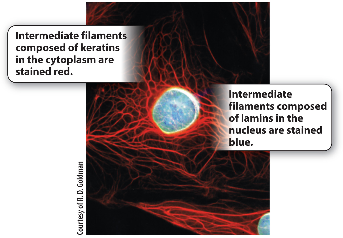
We have seen that different cell types use the same tubulin dimers to form microtubules and the same actin monomers to form microfilaments. By contrast, the proteins making up intermediate filaments differ from one cell type to another. For example, in epithelial cells, these protein subunits are keratins; in fibroblasts, they are vimentins; and in neurons, they are neurofilaments. Some intermediate filaments, called lamins, are even found inside the nucleus, where they provide support for the nuclear envelope (Fig. 10.10). There are well over 100 different kinds of intermediate filaments.
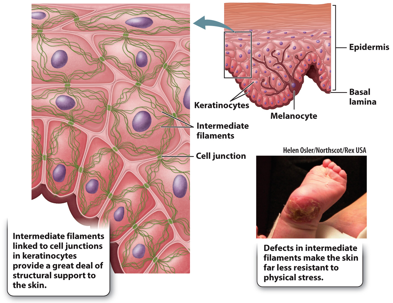
Once assembled, many intermediate filaments become attached to cell junctions at their cytoplasmic side, providing strong support for the cells (Fig. 10.11). In the case of epithelial cells, this anchoring results in structural continuity from one cell to another that greatly strengthens the entire epithelial tissue. This is especially important for tissues that are regularly subject to physical stress, such as the skin and the lining of the intestine.
205
Genetic defects that disrupt the intermediate filament network can have severe consequences. For example, some individuals with epidermolysis bullosa, a group of rare genetic diseases, have defective keratin genes. Intermediate filaments do not polymerize properly in these individuals, thus forming weaker connections between the layers of cells that make up the epidermis. As a consequence, the outer layers can detach, resulting in extremely fragile skin that blisters in response to the slightest trauma (Fig. 10.11). The sensitivity to physical stress is so extreme that infants with epidermolysis bullosa often suffer significant damage to the skin during childbirth. Therefore, Caesarean section is sometimes recommended in cases where the disease is diagnosed during pregnancy.