Prophase: Chromosomes condense and become visible.
Mitosis takes place in five stages, each of which is easily identified by events that can be observed in the microscope (Fig. 11.5). When you look in a microscope at a cell in interphase, specific chromosomes cannot be distinguished because they are long and thin. As the cell moves from G2 phase to the start of mitosis, the chromosomes condense and become visible in the nucleus. The first stage of mitosis is known as prophase and is characterized by the appearance of visible chromosomes.
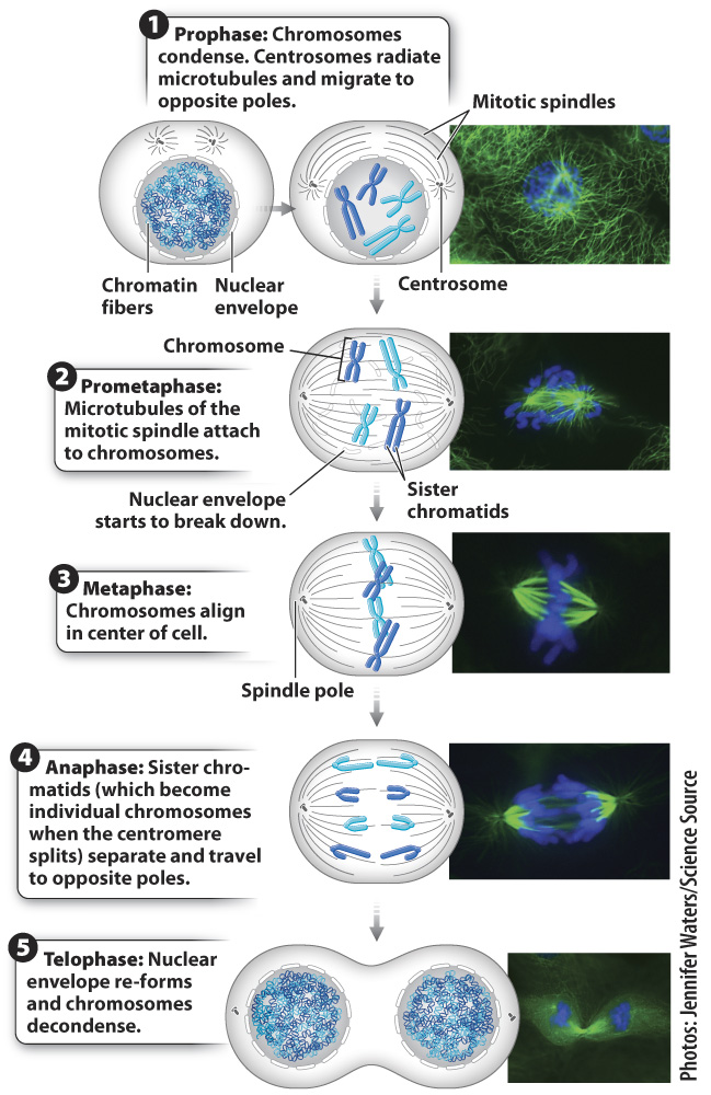
Outside the nucleus, in the cytosol, the cell begins to assemble the mitotic spindle, a structure made up predominantly of microtubules that pull the chromosomes into separate daughter cells. Recall from Chapter 10 that the centrosome is a compact structure that is the microtubule organizing center for animal cells. The centrosome is thus the structure from which the spindles radiate. Plant cells also have microtubule-
As part of the preparation for mitosis during S phase in animal cells, the centrosome duplicates and each one begins to migrate around the nucleus, the two ultimately halting at opposite poles in the cell at the start of prophase. The final locations of the centrosomes define the opposite ends of the cell that will eventually be separated into two daughter cells. As the centrosomes make their way to the poles of the cell, tubulin dimers assemble around them, forming microtubules that radiate from each centrosome. These radiating filaments form the mitotic spindle and later serve as the guide wires for chromosome movement.
224