DNA sequencing makes use of the principles of DNA replication.
The ability to determine the nucleotide sequence of DNA molecules has given a tremendous boost to biological research. Techniques used to sequence DNA follow from our understanding of DNA replication. Consider a solution containing identical single-
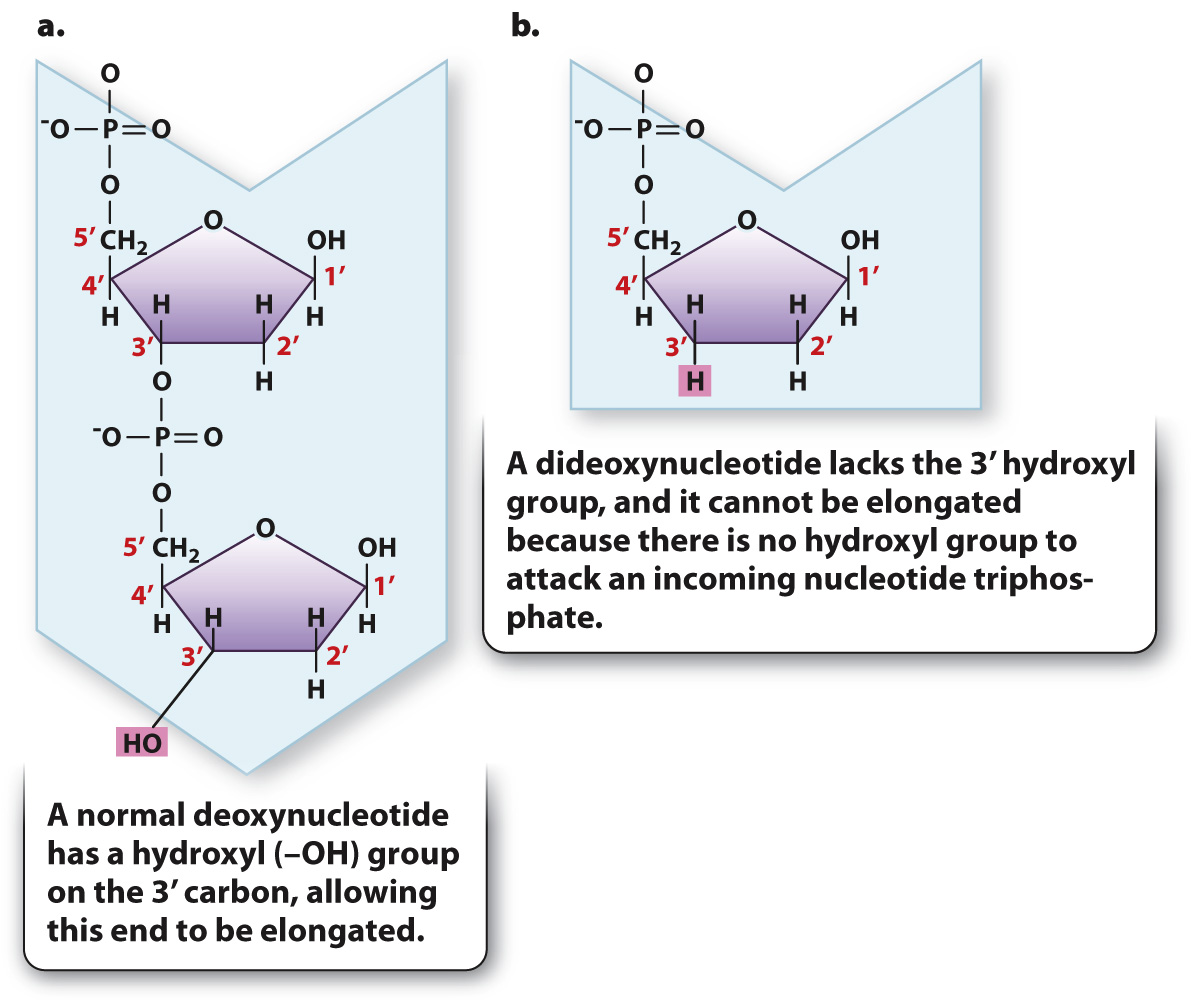
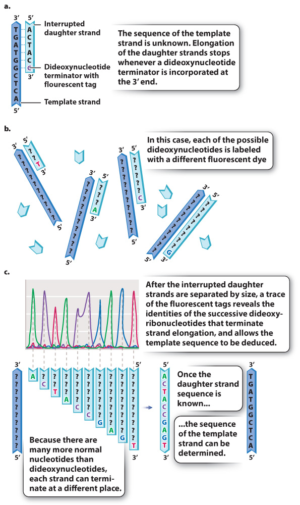
Recall that a free 3′ hydroxyl group is essential for each step in elongation because that is where the incoming nucleotide is attached (Fig. 12.17a). Making use of this fact, Sanger synthesized dideoxynucleotides, in which the 3′ hydroxyl group on the sugar ring is absent (Fig. 12.17b). Whenever a dideoxynucleotide is incorporated into a growing daughter strand, there is no hydroxyl group to attack the incoming nucleotide, and strand growth is stopped dead in its tracks (Fig. 12.18a). For this reason, a dideoxynucleotide is known as a chain terminator. By including a small amount of each of the chain terminators in a reaction tube along with larger quantities of all four normal nucleotides, a DNA primer, a DNA template, and DNA polymerase, Sanger was able to produce a series of interrupted daughter strands, each terminating at the site at which a dideoxynucleotide was incorporated.

Fig. 12.18b shows how the interrupted daughter strands help us to determine the DNA sequence by the procedure now called Sanger sequencing. In a tube containing dideoxy-

264
Each of the four dideoxynucleotides is chemically labeled with a different fluorescent dye, as indicated by the different colors of A, C, T, and G in Fig. 12.18b, and so all four terminators can be present in a single reaction and still be distinguished. After DNA synthesis is complete, the daughter strands are separated by size with gel electrophoresis. The smallest daughter molecules migrate most quickly and therefore are the first to reach the bottom of the gel, followed by the others in order of increasing size. A fluorescence detector at the bottom of the gel “reads” the colors of the fragments as they exit the gel. What the scientist sees is a trace (or graph) of the fluorescence intensities, such as the one shown in Fig. 12.18c. The differently colored peaks, from left to right, represent the order of fluorescently tagged DNA fragments emerging from the gel. Thus, a trace showing peaks colored green-
Quick Check 3 You have determined that the newly synthesized strand of DNA in your sequencing reaction has the sequence 5′-ACTACCGAGT-
Quick Check 3 Answer
The sequence of the template strand is antiparallel to the synthesized strand and inferred from complementary base pairing as 5′-ACTCGGTAGT-