A signaling molecule can cause multiple responses in the cell.

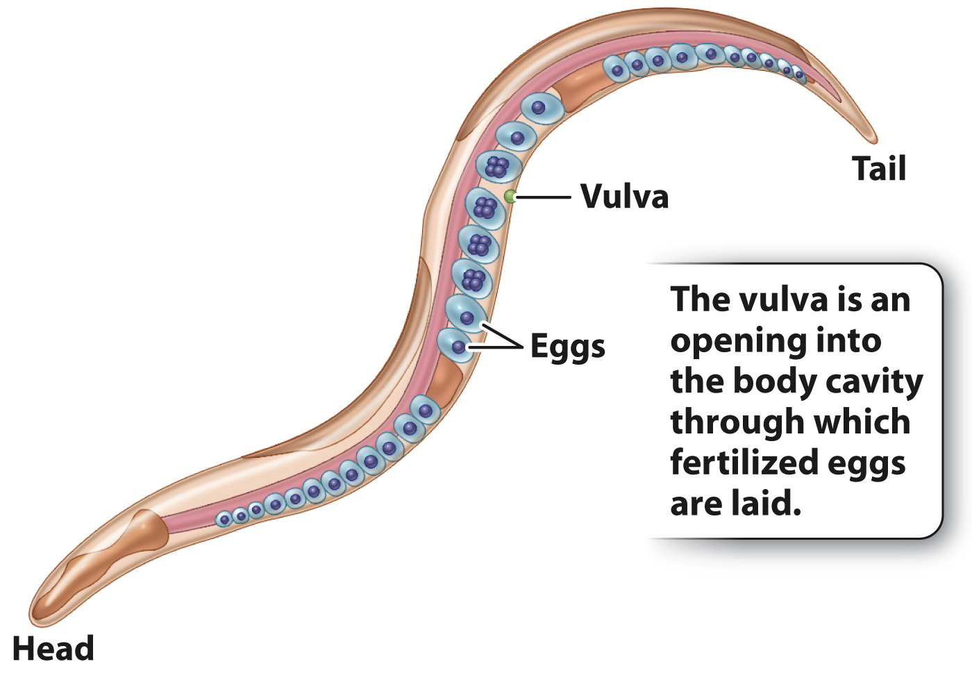
Pioneering experiments on signal transduction in development were carried out in the soil nematode Caenorhabditis elegans (Fig. 20.23). This worm is a useful model organism for developmental studies because of its small size (the adult is about 1 mm long), short generation time (2½ days from egg to sexually mature adult), and stereotyped process of development. (A stereotyped process is one that is always the same in each individual.) The adult animal consists of exactly 959 somatic (body) cells, and the cells’ patterns of division, migration, differentiation, and morphogenesis are identical in each individual C. elegans. The adult is typically a hermaphrodite that produces both male and female reproductive cells. The eggs are fertilized internally by sperm, and after undergoing five or six cell divisions the eggs are laid through an organ known as the vulva (Fig. 20.23).
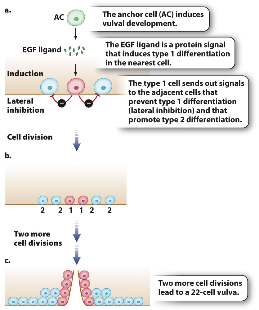
Vulva development is relatively simple and therefore amenable to experimental studies. The entire structure arises from only three multipotent cells that undergo either of two types of differentiation and divide to produce a vulva consisting of exactly 22 cells. Cells resulting from type 1 differentiation form the opening of the vulva, and cells resulting from type 2 differentiation form a supporting structure around the vulval opening. In each of the two types of differentiation, different genes are activated or repressed, resulting in differentiated cells with different functions.
How each of the three original cells, called progenitor cells, differentiates is determined by its position in the developing worm (Fig. 20.24). The fate of the progenitor cells is determined by their proximity to another cell, called the anchor cell (“AC” in Fig. 20.24a), which secretes a protein called epidermal growth factor (EGF) that binds to and activates a transmembrane EGF receptor (Chapter 9). The progenitor cell closest to the anchor cell receives the most amount of signal, and upon activation of its EGF receptor the progenitor cell carries out three functions:
416
It activates the genes for differentiation into a type 1 cell (Fig. 20.24a).
It prevents the adjacent cells from differentiating as type 1 (a process called lateral inhibition).
It induces the adjacent cells to differentiate as type 2 cells.
