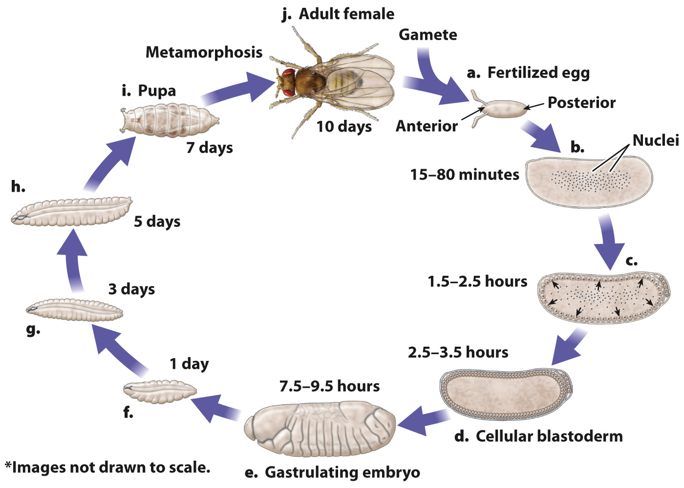
Drosophila development proceeds through egg, larval, and adult stages.
The fruit fly Drosophila melanogaster has played a prominent role in our understanding of the genetic control of early development, and in particular the hierarchical control of development. Researchers have isolated and analyzed a large number of mutant genes that lead to a variety of defects at different stages in development. These studies have revealed many of the key genes and processes in development, which are the focus of the following sections.
The major events in Drosophila development are illustrated in Fig. 20.5. DNA replication and nuclear division begin soon after the egg and sperm nuclei fuse (Fig. 20.5a). Unlike in mammalian development, the early nuclear divisions in the Drosophila embryo occur without cell division, and therefore the embryo consists of a single cell with many nuclei in the center (Fig. 20.5b). When there are roughly 5000 nuclei, they migrate to the periphery (Fig. 20.5c), where each nucleus becomes enclosed in its own cell membrane, and together they form the cellular blastoderm (Fig. 20.5d).

Then begins the process of gastrulation, in which the cells of the blastoderm migrate inward, creating layers of cells within the embryo. As in humans and most other animals (section 20.1 and Chapter 42), gastrulation forms the three germ layers (ectoderm, mesoderm, and endoderm) that differentiate into different types of cell. A Drosophila embryo during gastrulation is shown in Fig. 20.5e. At this stage, the embryo already shows an organization into discrete parts or segments, the formation of which is known as segmentation. There are three cephalic segments, C1–
About one day after fertilization, the embryo hatches from the egg as a larva (Fig. 20.5f). Over the next eight days, the larva grows and replaces its rigid outer shell, or cuticle, twice (Fig. 20.5g and 20.5h). After a week of further growth, the cuticle forms a casing—
405