The resting membrane potential is negative and results in part from the movement of potassium ions.
The inside surface of a neuron carries an electrical charge that is due to the presence of charged ions, and the same is true of the outside surface. However, the electrical charge on the inside and the electrical charge on the outside are not the same because unequal numbers of charged ions are located inside and outside the cell. When no signal is present, there are more negative ions inside the cell, and the inside is thus negatively charged relative to the outside.
The charge difference between the inside and the outside of a neuron due to differences in charged ions is the cell’s membrane potential, measured in volts. Other cells also have membrane potentials, but only nerve cells and muscle cells respond to changes in their membrane potential. Therefore, these cells are considered electrically excitable.
The key to producing an electrical signal in a nerve cell is the movement of positively and negatively charged ions across the cell membrane. This movement creates a change in electrical charge across the membrane that constitutes the electrical signal.
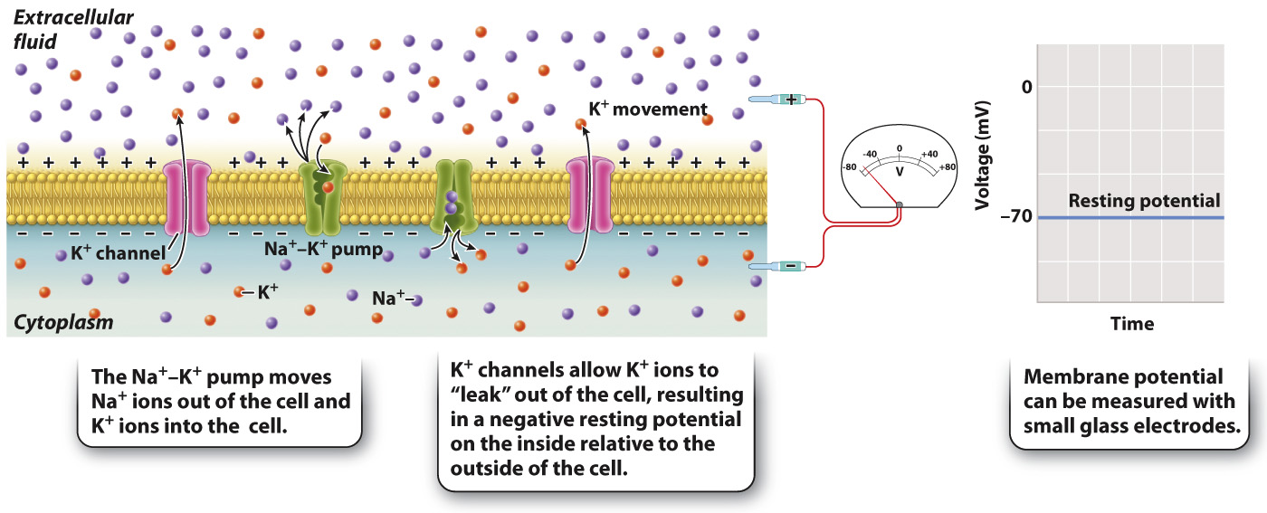
Let’s first consider the membrane potential when the neuron is at rest and no signal is being received or sent. Under these conditions, the cell’s membrane voltage is negative on its inside relative to its outside (Fig. 35.7). The resting membrane potential of the cell is said to be polarized. This means that there is a buildup of negatively charged ions on the inside surface of the cell’s plasma membrane and of positively charged ions on its outer surface. The negative voltage across the membrane at rest is referred to as the cell’s resting membrane potential The resting membrane potential ranges from –40 to –85 millivolts (mV) depending on the type of nerve cell and most commonly is about –65 to –70 mV. The voltage of the cell’s interior can be measured with respect to its outside voltage by small glass electrodes on the inside and outside of the cell.
746
At rest, nerve (and muscle) cells have a greater concentration of sodium (Na+) ions outside the cell than inside, and a greater concentration of potassium (K+) ions inside the cell than outside. This distribution of ions results in part from the action of the sodium-
The exact value of the resting membrane potential, however, depends on the movement of K+ ions back out of the cell by passive diffusion through potassium ion channels (Fig. 35.7). As discussed in Chapter 5, ion channels are protein pores embedded in the cell membrane. The most important ions that move across the cell membrane are sodium (Na+), potassium (K+), chloride (Cl−), and calcium (Ca2+). When a nerve cell is at rest, more K+ ion channels are open, giving K+ greater permeability compared with all other ions. As a result, K+ ions move out of the cell. The movement of K+ ions from the inside to the outside causes positive ions to build up on the outside of the cell, whereas negatively charged ions (largely proteins) remain on the inside of the cell, making the inside of the nerve cell more negative than its outside.
The relative proportion of ions does not by itself determine the resting membrane potential. This is because the number of charged ions that build up at the cell’s surface is a tiny fraction of the total number of charged ions and proteins located inside and outside the cell. It is the movement of K+ ions relative to other ions, particularly Na+ ions, that largely determines the resting membrane potential.