Signals between neurons can be excitatory or inhibitory.
Neurotransmitters bound to postsynaptic membrane receptors can either stimulate or inhibit the firing of action potentials in the postsynaptic neuron. The binding of some neurotransmitters depolarizes the postsynaptic membrane by opening ligand-
In contrast, the binding of other neurotransmitters can hyperpolarize the postsynaptic membrane, making the membrane potential more negative through the opening of ligand-
Excitatory synapses tend to transmit relevant information between nerve cells, and inhibitory synapses often serve to filter out unimportant information. For example, when a grazing gazelle senses a predator, its nervous system must transmit the key sensory information about the presence of a predator but filter out irrelevant information such as the flies buzzing over its back and ears. Inhibitory synapses are also important for controlling the timing of muscle activity required for coordinated movement, discussed in Chapter 37.
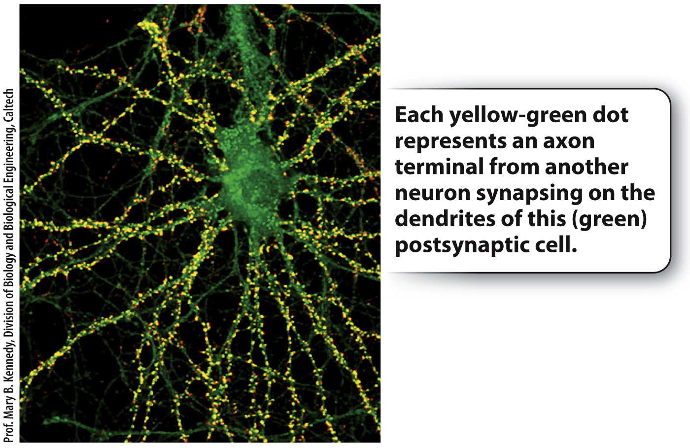
There are a number of different neurotransmitters, but a single nerve cell releases only one type of neurotransmitter. Consequently, a particular nerve cell exerts either an excitatory or an inhibitory effect on neighboring cells, but not both. In contrast, postsynaptic membranes typically contain multiple types of membrane receptor that bind different neurotransmitters. Thus, postsynaptic cells can receive both excitatory and inhibitory signals. Commonly, the dendrites or cell body of a single postsynaptic nerve cell make connections to hundreds or even thousands of axons from other nerve cells (Fig. 35.13).
The postsynaptic nerve cell sums the excitatory and inhibitory synaptic inputs (the summed EPSPs and IPSPs) that it receives through its dendrites and cell body (Fig. 35.14). If the sum of synaptic stimulation results in a membrane potential that exceeds threshold at the axon hillock, the neuron fires an action potential, communicating an impulse to cells that it in turn contacts. If the sum of inputs does not exceed threshold, no action potential fires (Fig. 35.14a).
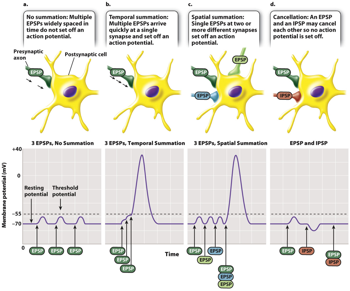
EPSPs and IPSPs can be summed over time and space. When summed over time, the frequency of synaptic stimuli determines whether the postsynaptic cell fires an action potential (Fig. 35.14b). This is referred to as temporal summation. When summed over space, the number of synaptic stimuli received from different regions of the postsynaptic cell’s dendrites determines if the cell fires an action potential (Fig. 35.14c). This is referred to as spatial summation. Sometimes, excitatory and inhibitory signals may cancel each other out (Fig. 35.14d). Temporal and spatial summation of EPSPs and IPSPs are the fundamental forms of information processing carried out by the nervous system. They provide mechanisms for determining whether a particular sensory stimulus is responded to or ignored. The role of temporal and spatial summation is explored further in Chapter 36.
753
More than 25 neurotransmitters are now recognized, and more are likely to be discovered. The amino acids glutamate (excitatory), glycine (inhibitory), and GABA (inhibitory) are key neurotransmitters that operate at synapses in the brain. The related amino acid derivatives dopamine, norepinephrine, and serotonin are also neurotransmitters acting in the brain. Simple peptides serve as neurotransmitters for sensory neurons. More recently, two gases, nitrous oxide and carbon monoxide, have been discovered to function as messengers between some nerve cells even though they do not behave as standard neurotransmitters that bind to membrane receptors.
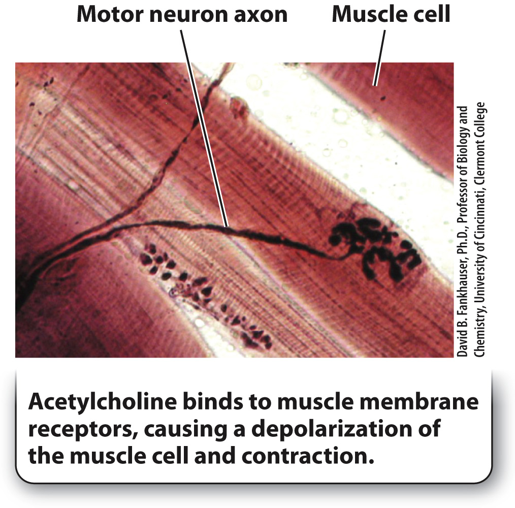
Acetylcholine is one of the key neurotransmitters produced in a variety of nerve cells. It is the excitatory neurotransmitter released by motor neurons to stimulate muscle fibers (Fig. 35.15). All vertebrate motor synapses rely on acetylcholine and are therefore excitatory only. The release of acetylcholine produces a depolarization of the muscle cell membrane by opening ligand-
Quick Check 4 Acetylcholinesterase is the enzyme that breaks down and inactivates acetylcholine. Some nerve gases used as chemical weapons block acetylcholinesterase. What effect would such nerve gases have on muscle contraction?
Quick Check 4 Answer
Nerve gases that block acetylcholinesterase, the enzyme that inactivates acetylcholine, cause violent muscle contractions, leading to paralysis of the diaphragm (the main muscle involved in breathing) and death by asphyxiation.