Chapter 35 Summary
Core Concepts Summary
35.1 Animal nervous systems allow organisms to sense and respond to the environment, coordinate movement, and regulate internal functions of the body.
Nerve cells, or neurons, receive and send signals and are the functional unit of the nervous system. page 740
Animal nervous systems include three types of neuron: sensory neurons that respond to signals, interneurons that integrate and process sensory information, and motor neurons that produce a response from muscle. page 740
Ganglia are localized collections of nerve cell bodies that integrate and process information. page 741
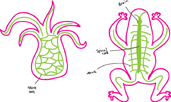
Simply organized animals, such as cnidarians, have a nerve net to coordinate sensory and motor function. page 741
Forward locomotion led to the evolution of specialized sense organs in the head, along with concentrated groupings of nerve cells to form ganglia and a brain. page 741
35.2 The basic functional unit of the nervous system is the neuron, which has dendrites that receive information and axons that transmit information.
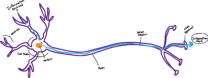
Neurons share a common organization: They have dendrites that receive inputs, a cell body that receives and sums the inputs, and axons that transmit signals to other nerve cells. page 743
Most neurons communicate by chemical synapses formed between an axon terminal and a neighboring nerve or muscle cell. page 743
Glial cells provide nutritional and physical support for neurons. page 744
35.3 The electrical properties of neurons allow them to communicate rapidly with one another.
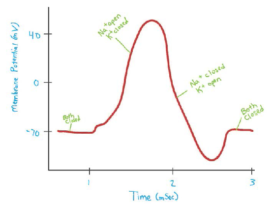
Neurons have electrically excitable membranes that code information by changes in membrane voltage and transmit information in the form of electrical signals called action potentials. page 745
Ion channels open and close in response to changes in membrane voltage, underlying the production of action potentials in nerve cells. page 746
Action potentials fire in an all-
In vertebrates, glial cells also produce the myelin sheath that insulates axons, increasing the speed of nerve impulses. page 749
Action potentials are conducted in a saltatory fashion in myelinated axons, firing at nodes of Ranvier, where the axon membrane is exposed and not insulated by myelin. page 749
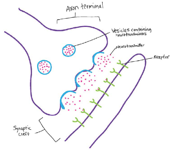
Communication across the synapse occurs when an arriving action potential triggers the release of neurotransmitters from vesicles within the axon terminal. page 751
Neurotransmitters released from presynaptic vesicles bind to receptors in the postsynaptic membrane, causing either an excitatory or an inhibitory stimulus. page 752
Excitatory stimuli depolarize the membrane, producing an excitatory postsynaptic potential (EPSP), whereas inhibitory stimuli hyperpolarize the membrane, producing an inhibitory postsynaptic potential (IPSP). page 752
760
35.4 Animal nervous systems can be organized into central and peripheral components.
Animal nervous systems are organized into central and peripheral components called the central nervous system (CNS) and peripheral nervous system (PNS). page 754
The central nervous system includes the brain and one or more main trunks of nerve cells, such as the spinal cord. The peripheral nervous system is distributed throughout the animal’s body and is composed of sensory and motor nerve cells. page 754
In many invertebrates and vertebrates, the peripheral nervous system is divided into voluntary and involuntary components. page 755
In vertebrates, the voluntary component is referred to as the somatic nervous system, and the involuntary component is referred to as the autonomic nervous system. page 755
The autonomic system regulates body functions through opposing actions of the sympathetic and parasympathetic divisions. page 756
The nervous system helps to regulate physiological functions to actively maintain stable conditions inside a cell or an organism, a process known as homeostasis. page 756
Homeostasis is often achieved by negative feedback, in which the response inhibits the stimulus. page 757
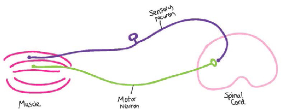
Simple reflex circuits can involve as few as two neurons: a sensory neuron from the periphery that synapses with a motor neuron in the spinal cord that sends a signal to a muscle. page 757
Self-Assessment
Diagram a simple nervous system of an animal that lacks cephalization and compare that system with the general organizational features of a nervous system that exhibits cephalization.
Self-Assessment 1 Answer
Name the three basic types of neuron and describe their functions.
Self-Assessment 2 Answer
Three basic categories of neuron are: (1) sensory neurons, which respond to physical features such as temperature, light, and touch, or to chemical signals such as molecules conveying odor and taste; (2) interneurons, which process the information received by the sensory neurons and transmit it to different body regions; and (3) motor neurons, which are at the end of the pathway and produce a suitable motor response based on the information from the sensory neurons and interneurons.
Diagram and label the basic features of a neuron, indicating where information is received and where it is sent.
Self-Assessment 3 Answer
Graph an action potential, showing the change in electrical potential on the y-
axis and time on the x- axis. Indicate on the graph the phases when voltage- gated Na+ and K+ ion channels are opened and when they are closed. Self-Assessment 4 Answer
Explain what is meant by saying action potentials are “all-
or- nothing.” Self-Assessment 5 Answer
An action potential is said to be “all-
or- nothing” because the magnitude of the action potential is always the same, regardless of the strength of the initial stimulating input. Explain why action potentials propagate along an axon only in a single direction.
Self-Assessment 6 Answer
A neuron cannot fire a second action potential during the refractory period, when the inner membrane voltage falls below and then returns to resting potential. The refractory period prevents the membrane from reaching its threshold too quickly after an action potential and firing an action potential in the reverse direction.
Create a table that lists the sequence of events that create an action potential, identifying the membrane potential at each stage, which channels are opening or closing, and in which direction ions are moving across the cell membrane.
Self-Assessment 7 Answer
Below is a sample table of action potential events:
Event
Membrane Potential
Channel
Ions
Resting membrane potential
‒70 mv
Stimulation causes EPSPs to be summed in dendrites and at cell body.
‒70 to ‒50 mv
Na+ channels open.
Na+ moves outside to inside.
Threshold potential reached.
‒50 mv
Na+ channels continue to open.
More Na+ moves outside to inside.
Action potential spike: Voltage rises
‒50 to +40 mv
More Na+ channels rapidly open.
Slow K+ channels begin to open.
More Na+ ions rush inside cell.
A few K+ ions move inside to outside
Action potential repolarization: Voltage falls
+40 to ‒80 mv
Na+ channels close.
K+ channels continue to open.
K+ moves inside to outside.
Refractory period
‒90 to ‒70 mv
K+ channels close.
Na+- K+ ATPase pumps active.
Na+ moves out.
K+ moves into cell.
Resting membrane potential re-
established. ‒70 mv
Na+ and K+ channels open and close at low rates.
A few more open K+ channels maintain negative resting membrane potential.
No net movement of ions across cell membrane.
Briefly describe how myelinated axons increase the speed of signal transmission.
Self-Assessment 8 Answer
The myelin sheath insulates the axon, spreading the charge from a local action potential over a much greater distance along the axon’s length. The axon membrane is exposed at regular intervals at sites called nodes of Ranvier that lie between adjacent segments wrapped with myelin. The action potential jumps from node to node, greatly increasing the speed of transmission.
Diagram a chemical synapse, labeling the vesicles that contain neurotransmitter molecules and the receptors that bind the neurotransmitter to produce either an inhibitory or an excitatory stimulus in the postsynaptic cell.
Self-Assessment 9 Answer
Describe how neurotransmitter binding to receptors on a postsynaptic cell causes inhibition or excitation.
Self-Assessment 10 Answer
Neurotransmitters binding to receptors on the postsynaptic membrane can elicit an excitatory response if they depolarize the postsynaptic membrane. Excitatory neurotransmitters trigger the opening of Na+ channels. Neurotransmitters cause an inhibitory response if they bind to receptors and hyperpolarize the postsynaptic membrane. Inhibitory neurotransmitters trigger the opening of Cl−, or sometimes K+, channels.
Describe how temporal summation of EPSPs from two presynaptic neurons results in a larger depolarization of the postsynaptic cell compared with how an inhibitory presynaptic neuron causes an IPSP that negates the EPSP of an excitatory neuron, and how spatial summation of three excitatory presynaptic neurons produces an even larger depolarization of the postsynaptic neuron.
Self-Assessment 11 Answer
Two EPSPs that arrive quickly one after the other are summed to produce a greater depolarization than either could alone. More Na+ ions diffuse into the cell through opened Na+ channels, causing a greater depolarization as the membrane potential becomes less negative. An IPSP from an inhibitory neuron makes the membrane potential more negative, or hyperpolarized, through the opening of ligand-
gated Cl- or K+ channels, allowing Cl- to diffuse into the cell or K+ to diffuse out. The hyperpolarization in membrane potential from an IPSP counteracts the depolarization in membrane potential due to an EPSP. Three EPSPs arriving at the cell at the same time at different regions of the postsynaptic cell’s dendrites produce an even larger depolarization of the postsynaptic neuron because more Na+ channels are opened simultaneously. List which functions of an animal are controlled by voluntary and by involuntary components of the nervous system.
Self-Assessment 12 Answer
The voluntary, or somatic, component of the vertebrate nervous system controls the sensing and response to external stimuli, like sight or smell. The involuntary, or autonomic, component of the vertebrate nervous system controls many bodily functions like heartbeat and digestion.
Diagram a simple circuit that includes a sensory neuron that synapses with a motor neuron to produce a reflex. Indicate where in the nervous system this synapse is found.
Self-Assessment 13 Answer