The structure and function of the vertebrate eye underlie image processing.
Vertebrate eyes are protected within the eye socket of the skull. The anatomy of the eye is shown in Fig. 36.14. The eye is surrounded by the sclera, a tough, white outer layer. Below the sclera, a thinner layer, called the choroid, carries blood vessels to nourish the eye. Mucus secretions produced over the sclera keep the eye moist. The portion of the sclera in the front of the eye is the cornea, which is transparent. Light passes through the cornea, enters the eye through an opening called the pupil, and then passes through a convex lens. Small muscles called ciliary muscles attach between the sclera and lens. Contraction or relaxation of these muscles adjusts the shape of the lens to focus light images.
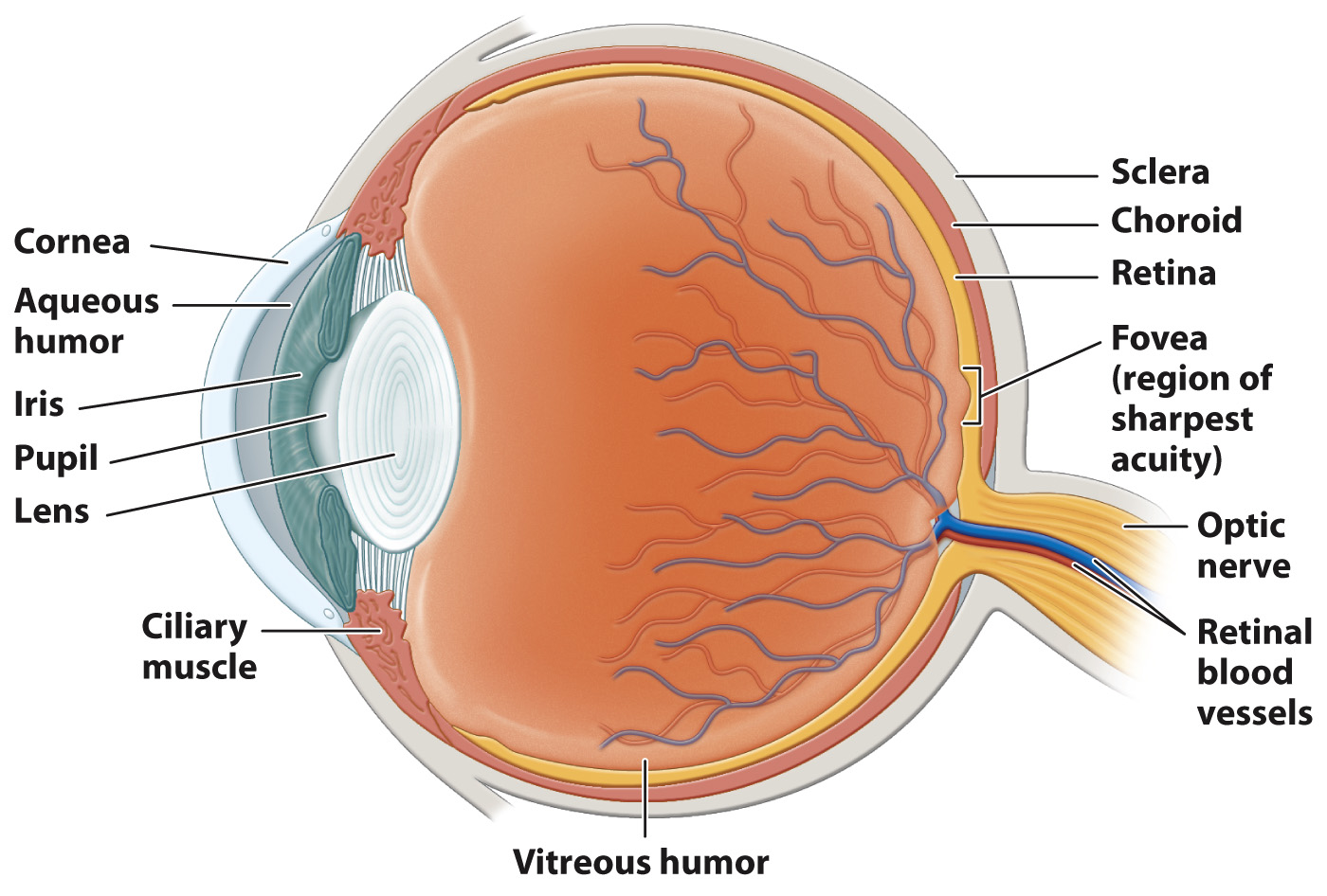
At the front of the eye, surrounding the pupil, the choroid forms a donut-
The focusing muscles and lens separate the eye into two regions. The interior region in front of the lens is filled with a clear watery liquid, the aqueous humor. This liquid is produced continuously and drained by small ducts at the base of the eye. The large cavity behind the lens is filled with a gel-
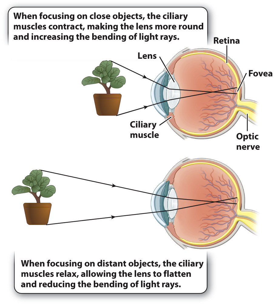
To form an image, the cornea and lens bend incoming light rays, focusing them on the retina (Fig. 36.14). In doing so, the light rays cross, inverting the image on the retina and changing up to down and left to right. The retina is a thin tissue located in the posterior of the eye. It contains the photoreceptors and nerve cells that sense and initially process light stimuli. Humans, other mammals, and birds focus light on the retina by changing the shape of the lens (Fig. 36.15). When we focus on objects at close range, as when we read a book, the ciliary muscles contract, making the lens more rounded to bend the light rays more. When we focus on distant objects, the ciliary muscles relax, allowing the lens to flatten, reducing the bending of light. In contrast, the eyes of octopus and squid focus by moving the lens back and forth within the eye, as a camera does, rather than by changing its shape.
774
Why do so many animals have two eyes? One reason is that two eyes provide a wider field of vision. Close one eye and you will see the visual field of the other eye. With both eyes open, the visual field is wider than the visual field of either eye alone. You’ll also notice considerable overlap in the visual field of both eyes. This overlap exists in humans and other primates, as well as in other mammals and birds of prey, and makes possible binocular vision, the ability to combine images from both eyes to produce a single visual image. Binocular vision allows for depth and distance perception.
Prey animals, such as some birds, antelope, and rabbits, have eyes positioned more to the side, expanding their visual field up to 360°, but limiting or preventing binocular vision. Animals that require greater visual sharpness, such as predatory hawks, cats, and snakes, have eyes pointing forward, increasing their binocular field of view.